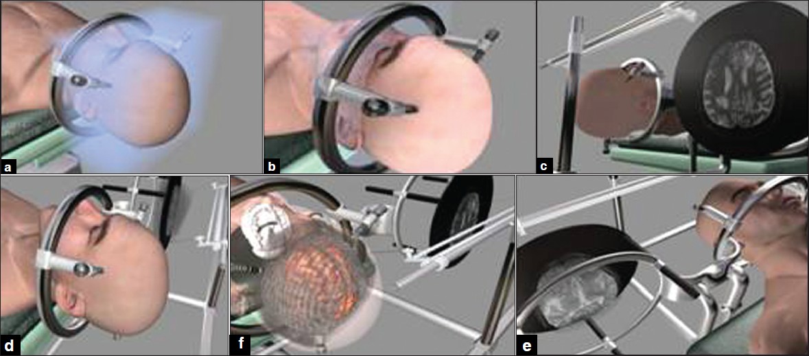
Figure 2: A right frontal oligodendroglioma confi rmed by stereotactic biopsy. (a) The frame has been fi xed to the cranium. (b) The head of patient have been fi xed at supine position to operating table and the MRI images have been transferred to monitor. (c and d) The patient and images on LCD data have been adjusted. (e and f) The pantoghraph show that the data of the patient head and pathology are seen at the same location of their counterpart on LCD.