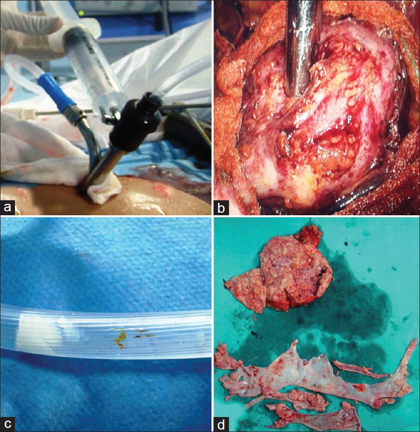
Figure 3: (a) Palanivelu hydatid system outer cannula consisting of two side channels for suction and irrigation. (b) Intraoperative placement of PHS over the surface of hydatid cyst. (c) Hydatid scolices seen through suction tube. (d) Renal hydatid cyst cut opened after surgical removal with aspiratedscolices and laminated membranes