
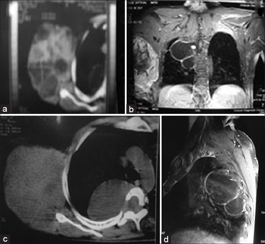
Figure 3: (a) CT scan of the tumor. (b) MRI showing both the tumor and the meningoceles. (c) CT scan showing lateral meningocele with a spinal defect and the tumor. (d) MRI lateral view showing the meningoceles
| Close | |
|
|
|
|
Figure 3: (a) CT scan of the tumor. (b) MRI showing both the tumor and the meningoceles. (c) CT scan showing lateral meningocele with a spinal defect and the tumor. (d) MRI lateral view showing the meningoceles
|
|