
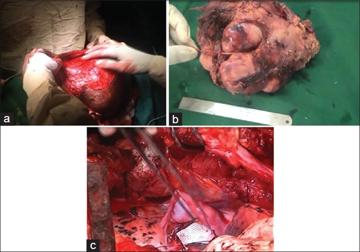
Figure 4: (a) Intraoperative photo while raising skin flaps. (b) Tumor with the lateral thoracic nerve excised in TOTO. (c) Repair of meningocele defects being done with a G‑patch (artificial dural patch)
| Close | |
|
|
|
|
Figure 4: (a) Intraoperative photo while raising skin flaps. (b) Tumor with the lateral thoracic nerve excised in TOTO. (c) Repair of meningocele defects being done with a G‑patch (artificial dural patch)
|
|