
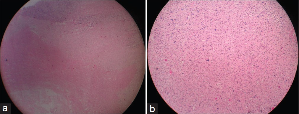
Figure 5: (a) Histopathology scanner view (×4) showing tumor with areas of necrosis. (b) Histopathology photographic view (×10) showing tumor cells arranged in fascicles with nuclear pleomorphism
| Close | |
|
|
|
|
Figure 5: (a) Histopathology scanner view (×4) showing tumor with areas of necrosis. (b) Histopathology photographic view (×10) showing tumor cells arranged in fascicles with nuclear pleomorphism
|
|