
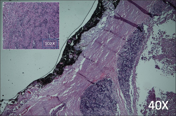
Figure 3: Photomicrograph shows encapsulated tumor with extensive necrosis with few preserved areas (H and E, ×40). High-power (inset) shows admixture of epithelial cells and small lymphocytes (H and E, ×100)
| Close | |
|
|
|
|
Figure 3: Photomicrograph shows encapsulated tumor with extensive necrosis with few preserved areas (H and E, ×40). High-power (inset) shows admixture of epithelial cells and small lymphocytes (H and E, ×100)
|
|