|
 
 |
| CASE REPORT |
|
| Year : 2016 | Volume
: 11
| Issue : 4 | Page : 133-136 |
|
Fracture of medial pole of right condyle and symphysis of mandible in a 6-year-old male: A conservative approach
Karthik Shunmugavelu1, Kumaravel Subramaniam2
1 Department of Dentistry and Faciomaxillary Surgery, Kasthuri Multispeciality Hospital, Chennai, Tamil Nadu, India
2 Department of Dentistry and Faciomaxillary Surgery, K. P. Multispeciality Hospital, Chennai, Tamil Nadu, India
| Date of Web Publication | 16-Mar-2017 |
Correspondence Address:
Karthik Shunmugavelu
Department of Dentistry and Faciomaxillary Surgery, Kasthuri Multispeciality Hospital, Shanmugam Road, West Tambaram, Chennai - 600 045, Tamil Nadu
India
  | Check |
DOI: 10.4103/1858-5000.202356 
Much importance has to be emphasized on the fractures of mandibular condyle and symphysis in children due to the growth center status. Risk involving this region might lead to facial asymmetry and growth retardation. Most common etiology includes trauma, fall, sports, occupational hazard, and interpersonal violence. Moreover, in the mandible, condyle is the foremost anatomical site to fracture. In this presentation, we highlight a case of a 6-year-old male witnessed with a fracture of unilateral medial pole of the right condylar head and mandibular symphysis. Clinical examination revealed limited mouth opening, edema, and deviation of mandible to the right side. Computed facial tomography revealed fractured right condylar head. Treatment plan included conservative approaches such as closed reduction and nonrigid mandibular splint. The main concept of this approach is to emphasize that nonsurgical and functional approach plays a crucial role in the management of mandible symphysis and condyle fractures in pediatric population thereby avoiding growth inhibition. Keywords: Condyle, conservative approach, mandible, pediatric, trauma
How to cite this article:
Shunmugavelu K, Subramaniam K. Fracture of medial pole of right condyle and symphysis of mandible in a 6-year-old male: A conservative approach. Sudan Med Monit 2016;11:133-6 |
| Introduction | |  |
The mandible is the most commonly involved facial bone in trauma. Among the parts of the mandible, condyle is the most commonly involved anatomical site constituting 19%–52%.[1],[2] Head, neck, and subcondyle are the subdivisions of the condyle. Fracture of condyle in the pediatric age group is usually witnessed as greenstick type rather than displaced.[3],[4] Etiology includes trauma, fall, road traffic accidents, sports activities, occupational hazard, and interpersonal violence. Trauma to the condyle is to be considered seriously because of the mandibular growth center in the cartilage of the condyle head. Risk might lead to growth retardation, facial asymmetry, deviation of the mandible to the affected side, muscle spasm, restricted mouth opening, and pain. Regeneration of the condylar process is contributed by articular disc and capsule.[5],[6],[7] In this case report, we highlight a challenging clinical situation of fracture involving medial pole of the right condylar head and mandibular symphysis in a 6-year-old male who underwent closed reduction and nonrigid mandibular splint.
| Case Report | | |
A 6-year-old male attended to the Department of Dentistry and Faciomaxillary Surgery with swelling on the right side of face caused due to trauma. The patient was conscious, oriented, and afebrile. Vitals were stable. Clinical examination revealed edema, pain, restricted mouth opening, deviation of the mandible to the affected side, anterior crossbite, crowding, defective mastication, incorrect speech, facial asymmetry, and restricted lateral temporomandibular joint (TMJ) movements. Past medical and dental drug histories revealed no significance. Radiological investigations such as facial computed tomography–axial and sagittal sections–revealed fracture of medial pole of the right condylar head and mandibular symphysis [Figure 1] and [Figure 2].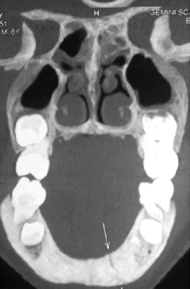 | Figure 1: Coronal facial computed tomography depicting fractured mandible symphysis (arrows)
Click here to view |
 | Figure 2: Sagittal facial computed tomography depicting fractured medial pole of the right condylar head
Click here to view |
Treatment
Two-stage treatment protocol was planned such that fibrous union and remodeling can occur simultaneously. The first priority was that of closed reduction of the fractured segments followed by insertion of a nonrigid mandibular splint which was prepared based on maxillary and mandibular impression. Cast models were created and finally fabrication of removable mandibular splint measuring 3–3.5 mm was done. This avoids direct force on the mandible thereby providing functional reposition. An effective reduction and remodeling was evident after 2-, 4-, 6-, 8-, 10-, and 12-month follow-up. Postoperative clinical findings include absence of pain, swelling, discomfort, stable occlusion, satisfactory mouth opening, and lateral TMJ movements. Postoperative complications such as ankylosis, TMJ dysfunction, and defective mouth opening were not seen. After complete and satisfactory healing of the fractured segments, further dental treatment procedures were planned.
| Discussion | | |
In trauma, mandible and condyle are the most commonly involved facial bone and anatomical sites, respectively. Etiology includes road traffic accidents, trauma, fall, sports activities, interpersonal violence, and occupational hazard. Various factors which influence the treatment plan include age of the patient, dentition, occlusion, level, displacement, comorbidities, and side–ipsilateral or contralateral.[2],[5] Clinical findings include edema on the affected side, deviation of the mandible to the affected side, deranged occlusion, restricted mouth opening and lateral TMJ movements, pain, tenderness, and facial asymmetry.[2],[5],[8] Based on the age group of the patient, treatment methods such as closed reduction or surgical approach have been planned. In children, closed reduction followed by intermaxillary fixation (IMF) is the treatment of choice. The main concept of this treatment is to focus on early mobilization, functional stimulation, and bone remodeling. Whereas, the surgical approach is indicated in adults with displaced or dislocated condylar heads, such as open reduction and internal fixation and IMF.[2],[6],[9] One main advantage in pediatric population is that rapid healing is observed within a span of 3–4 weeks compared to adults which might take 6 months. Very rarely, nonunion or fibrous union is seen in children. Long-term outcome aims at inhibition of growth disturbances followed by functional remodeling, usually attained by closed reduction method in the pediatric population. Internal factors such as remodeling of trabecular bone followed by endochondral ossification occur during the healing period of condylar fractures in children, thereby preserving the articular disc, fibrous capsule, and cartilage of the condyle.[7],[10]
| Conclusion | | |
In our case, nonsurgical approach was planned to promote active growth since the affected area was that of mandibular condylar head. This, in turn, will stimulate functional remodeling and facial symmetry through formation of new sites of bone during chondrocyte proliferation. Normal architecture of the condyle-glenoid fossa relationship is obtained through functional remodeling and adaptation. Finally, restoration of the esthetics, structure, and function, lost due to trauma, was achieved in a remarkable manner.
Financial support and sponsorship
Nil.
Conflicts of interest
There are no conflicts of interest.
| References | | |
| 1. | Tuna EB, Dündar A, Cankaya AB, Gençay K. Conservative approach to unilateral condylar fracture in a growing patient: A 2.5-year follow up. Open Dent J 2012;6:1-4.  |
| 2. | Zachariades N, Mezitis M, Mourouzis C, Papadakis D, Spanou A. Fractures of the mandibular condyle: A review of 466 cases. Literature review, reflections on treatment and proposals. J Craniomaxillofac Surg 2006;34:421-32. |
| 3. | Kalia V, Singh AP. Greenstick fracture of the mandible: A case report. J Indian Soc Pedod Prev Dent 2008;26:32-5. [ PUBMED] [Full text] |
| 4. | Li Z, Zhang W, Li ZB, Li JR. Mechanism in favorable prognosis of pediatric condylar fractures managed by closed procedures: An experimental study in growing rats. Dent Traumatol 2010;26:228-35. |
| 5. | Farronato G, Grillo ME, Giannini L, Farronato D, Maspero C. Long-term results of early condylar fracture correction: Case report. Dent Traumatol 2009;25:e37-42. |
| 6. | Valiati R, Ibrahim D, Abreu ME, Heitz C, de Oliveira RB, Pagnoncelli RM, et al. The treatment of condylar fractures: To open or not to open? A critical review of this controversy. Int J Med Sci 2008;5:313-8. |
| 7. | Hackett JF, Sleeman DJ. Vertical-split fracture of mandibular condyle and its sequelae. Br Dent J 2001;191:557-8. |
| 8. | Silvennoinen U, Lindqvist C, Oikarinen K. Dental injuries in association with mandibular condyle fractures. Endod Dent Traumatol 1993;9:254-9. |
| 9. | De Riu G, Gamba U, Anghinoni M, Sesenna E. A comparison of open and closed treatment of condylar fractures: A change in philosophy. Int J Oral Maxillofac Surg 2001;30:384-9. |
| 10. | Lloyd T, Nightingale C, Edler R. The use of vacuum-formed splints for temporary intermaxillary fixation in the management of unilateral condylar fractures. Br J Oral Maxillofac Surg 2001;39:301-3. |
[Figure 1], [Figure 2]
|


 Search Pubmed for