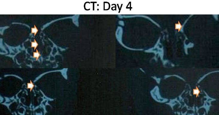
Figure 2: Coronal computed tomography bone scan of skull base for day 4 infant revealed that orbital plates of the frontal bone was completely ossified and had a thin medial end extend over the lateral ethmoidal cells. Roof of ethmoid complex was ossified partially with gap between the ossified medial and lateral extensions. Vomer was ossified, but not meet perpendicular plate of the ethmoid in posterior nasal septum. Crista galli was not been ossified yet, so it was not fused with the ethmoidal labyrinths. Ethmoidal air cells were developed. No extension above the plane of the cribriform plate. The cribriform plate was not ossified