|
|
Pathological Case of the Month
Sonya R. Arnold, MD;
Orestes Borrego, MD;
Enid Gilbert-Barness, MD
Arch Fam Med. 1998;7:513-514.
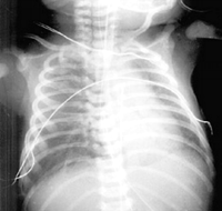
A male infant, weighing 2845 g, was born at 42 weeks' gestation to a 27-year-old, gravida 1, para 0 mother. The pregnancy was complicated by a group B streptococcal infection in the mother. Labor was induced and complicated by fetal distress. The infant was delivered by cesarean section amid thick meconium fluid. A chest x-ray film showed moderate lung disease and small bilateral infiltrates (Figure 1). The infant had multiple neurological symptoms, including a staring gaze, dilated pupils that were unresponsive to light, and lip smacking. There was no response to tactile stimuli, and deep tendon reflexes were absent in all extremities. The infant's condition continued to deteriorate, with a failed response to high-frequency ventilation. Arterial blood gas values were as follows: pH, 6.82; oxygen pressure, 12 mm Hg; carbon dioxide pressure, 98 mm Hg; oxygen saturation, 5%; base excess, -20 mEq/L; and bicarbonate, 10 mEq/L. Extracorporeal membrane oxygenation therapy was suggested, but the patient went into cardiac arrest and did not respond to aggressive attempts at resuscitation. The infant was pronounced dead 22 hours after birth.
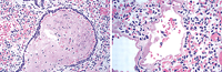
At autopsy, the lungs were atelectatic and hypocrepitant. Microscopic sections of the lungs are shown in Figure 2.
From the Department of Pathology, Tampa General Hospital, University of South Florida, Tampa.
|
Diagnosis and Discussion: Meconium Aspiration Syndrome
Figure 1. Chest x-ray film showing bilateral lung infiltrates.
Figure 2. Microscopic sections showing squamous cells and meconium (left) and hyaline-membrane formation (right) (hematoxylin-eosin, x100 [left] and x250 [right]).
Meconium is a green viscous fluid consisting of fetal gastrointestinal tract secretions. Meconium-stained amniotic fluid occurs in 12% to 13% of live births in the United States each year.1-2 Meconium aspiration is defined as meconium presence below the vocal cords.3-4 Studies show that meconium aspiration syndrome develops in 2% to 5% of infants with meconium-stained amniotic fluid.1-2 The syndrome is diagnosed when respiratory distress develops in an infant soon after birth in the presence of meconium staining and radiographic evidence of aspiration pneumonitis.3-4
Etiologic factors are not entirely clear. Meconium passage is associated with fetal maturity. A study by Wiswell and Bent2 revealed meconium passage in at least 35% of pregnancies extending beyond the 42nd week. Passage of meconium has also been associated with fetal asphyxia. One current hypothesis is that in utero hypoxia stimulates a vagal response resulting in increased peristalsis and relaxation of anal sphincter tone.2, 5 In the same way, fetal head compression or cord compression may cause a vagal response.
The mechanism of meconium aspiration syndrome remains a point of controversy. Katz and Bowes1 propose that aspiration occurs in utero when a distressed fetus begins gasping. They strongly associate the syndrome with asphyxia, which so severely damages the lungs that meconium and other aspirated material cannot be adequately cleared. Others2, 6-7 contend that aspiration occurs in the perinatal period just as the neonate takes the first breaths.
Aspirated meconium exerts what has been described by some authors as a ball-valve effect, ie, air flows into the airspaces on inspiration but is trapped in these spaces on expiration. This air-trapping can lead to overdistension of the distal airways, alveolar rupture, and air leaks.5, 8 An air leak can cause a pneumothorax or pneumomediastinum. Complete obstruction can lead to atelectasis and ventilation perfusion mismatch.5 With continued breathing, the aspirated meconium migrates peripherally.2, 8 Clinical manifestations may include tachypnea, retraction, grunting, and cyanosis.6 Likewise, these changes are often reflected in the chest x-ray films, which typically show bilateral infiltrates, pneumothoraces and/or pneumomediastina, and atelectasis1, 5; also, the diaphragm may be flattened.
Persistent pulmonary hypertension has been linked to meconium aspiration syndrome with a high mortality rate. In a study of 11 consecutive infants with fatal meconium aspiration, Murphy et al9 found evidence of persistent pulmonary hypertension in 10.
Histologic findings include excessive muscularization of the intra-acinar pulmonary arteries.9 These findings are also notable because muscularization of the arteries is a chronic change and the meconium aspiration syndrome occurs acutely. This suggests that meconium passage may be an indicator of intrauterine hypoxia.10
Frequent autopsy findings include a green discoloration of the lungs and meconium in the bronchi. Microscopic sections may show fetal squamous cells and mucus. Within hours, the alveoli can be filled with neutrophils and basophilic debris11 and muscularization of the pulmonary arteries may be seen in cases associated with persistent pulmonary hypertension. Hyaline membranes may also develop, and pulmonary hemorrhage and vascular necrosis may appear, depending on the degree of asphyxia.
Current therapy includes thorough catheter suctioning of secretions from the oropharynx before the infant takes his or her first breath.10 In depressed infants and those delivered amid thick meconium, endotracheal intubation with suctioning can be performed. Preventive measures include careful monitoring for fetal distress with prompt intervention. Extracorporeal membrane oxygenation has contributed to increased survival and may benefit patients who are refractory to mechanical ventilation.11
Prognosis depends on the extent of central nervous system damage secondary to asphyxia or other sequelae such as persistent fetal circulation.11 Studies12 have also shown an increased incidence of respiratory morbidity in infants during the first 6 months of life.
Selected from Arch Pediatr Adolesc Med. 1996;150:1217-1218. Pathological Case of the Month.
REFERENCES
|
1. Katz VL, Bowes WA. Meconium aspiration syndrome: reflections on a murky subject. Am J Obstet Gynecol. 1992;166:171-183.
WEB OF SCIENCE
| PUBMED
2. Wiswell TE, Bent RC. Meconium aspiration and the meconium aspiration syndrome: unresolved issues. Pediatr Clin North Am. 1993;40:955-981.
WEB OF SCIENCE
| PUBMED
3. Gautham KS, Narang A. Reply to meconium aspiration syndrome: recent concepts. Ind Pediatr. 1994;31:1001-1002.
4. Kumar A, Mishra PK. Meconium aspiration syndrome: recent concepts. Ind Pediatr. 1993;30:5-7.
5. Moore CS. Meconium aspiration syndrome. Neonat Network. 1994;13:57-60.
6. Behrman RE. Nelson Textbook of Pediatrics. 14th ed. Philadelphia, Pa: WB Saunders Co; 1992:469-470.
7. Cunningham FG, MacDonald PC, Leveno KJ, Gant NF, Gilstrap LC III. Williams Obstetrics. 19th ed. Baltimore, Md: Williams & Wilkins; 1993:995-996.
8. Gregory GA, Gooding CA, Phibbs RH, Tooley WH. Meconium aspiration in infants: a prospective study. J Pediatr. 1974;85:848-852.
FULL TEXT
|
WEB OF SCIENCE
| PUBMED
9. Murphy JD, Vawter GF, Reid LM. Pulmonary vascular disease in fatal meconium aspiration. J Pediatr. 1984;104:758-762.
WEB OF SCIENCE
| PUBMED
10. Cunningham AS, Lawson EE, Martin RJ, Pildes RS. Tracheal suction and meconium: a proposed standard of care. J Pediatr. 1990;116:153-154.
FULL TEXT
|
WEB OF SCIENCE
| PUBMED
11. Stocker JT, Dehner LP. Pediatric Pathology. Philadelphia, Pa: JB Lippincott Co; 1992:545.
12. Yuksel B, Greenough A, Gamsu HR. Neonatal meconium aspiration syndrome and respiratory morbidity during infancy. Pediatr Pulmonol. 1993;16:358-361.
PUBMED
|