|
|
An Ulcerating Nodule on the Arm
Contributor Malinee Saxena, BA;
Contributor Ellen B. Rest, MD;
Section Editor Lori Lowe, MD
Arch Fam Med. 1999;8:97.
A 27-year-old white woman presented to the student health clinic with a 2-week history of an enlarging ulcerated nodule on the dorsum of her left wrist. In the weeks preceding the visit, she had worked in her garden and had traveled to the West Indies. There was no history of trauma to the area, and she denied generalized symptoms. She was otherwise well. The lesion started as a pinpoint red papule that ulcerated as it expanded in depth and width. The erythematous surrounding tissue was mildly painful. Also, within the prior 2 weeks, the patient had noted the appearance of several palpable nodules on the upper part of her left arm.
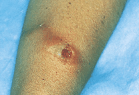
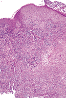
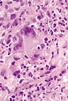
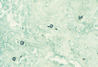
On examination, the patient was afebrile. A 2.5-cm-diameter nodule with a 1-cm deep ulceration was located on the dorsum of the left wrist. A curvilinear erythematous patch extended from the nodule radially and proximally (Figure 1). At the time of presentation, 2 firm subcutaneous nodules without overlying skin changes were palpable over the left biceps. Three biopsy specimens from the periphery of the nodule were obtained for culture and pathologic examination (Figure 2, Figure 3 and Figure 4).
What is your diagnosis?
From the University of Minnesota, Minneapolis.
|
Diagnosis and Discussion: Lymphocutaneous sporotrichosis.
HISTOPATHOLOGIC AND CULTURE RESULTS
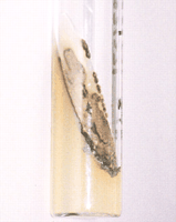
The biopsy specimens showed an area of central dermal necrosis surrounded by a mixed infiltrate of lymphocytes, histiocytes, plasma cells, and neutrophils. Occasional multinucleate giant cells were present. A Gomori methenamine-silver stain revealed yeast forms, some of which had a round or an elongated bud (Figure 4). The fungal culture yielded Sporothrix schenkii in a characteristic dark, leatherlike plaque (Figure 5).
DISCUSSION
Lymphocutaneous sporotrichosis, a granulomatous disease, was first described in 1898 by Schenck, who isolated the fungus S schenkii. Sporotrichosis appears worldwide but prefers temperate and tropical climates that support saprophytic growth. Although the infection may occur in all age groups, it is most common in adult men who are horticulturists, florists, farmers, gardeners, or veterinarians.1 Exposure to such plants as rosebushes, barberry, sphagnum moss, contaminated mine timbers, and other sharp vegetation2 or contact with such animals as horses, fish, birds, reptiles, cats, dogs, and rats is a possible source of sporotrichosis.3
Once the skin has been inoculated, a papule will form that progresses to form a pustule and then a nodule with ragged undetermined borders and a central ulcer. The nodule is mobile and not tender unless it is secondarily infected with bacteria. With lymphatic spread, more proximal subcutaneous nodules will appear in a linear fashion.4 The fungus tends to produce an insidious infection that is not invasive or virulent.2 If not treated, secondary sporotrichosis nodules may be gummatous and may persist for months to years.4
Although the majority of cases of sporotrichosis in the United States are lymphocutaneous, other forms, such as fixed cutaneous, extracutaneous, and disseminated, may occur. Fixed cutaneous lesions are most commonly found on the hands of adults and the faces of young; without lymphatic spread, the infection is localized to the site of infection. Patients with fixed cutaneous sporotrichosis usually live in endemic areas and have a high degree of immunity. The extracutaneous and disseminated forms of the disease are uncommon and occur in immunocompromised individuals. In these patients, exposure to plant matter may not precede the infection. Pulmonary infection results from inhalation or aspiration of the fungus, causing pneumonitis with fibrocavitary disease that progressively worsens.4
In most cases, the findings of histologic examination are unreliable in the diagnosis of sporotrichosis, as fungal elements are difficult to find in sporadic histologic sections; however, Japanese researchers have been able to demonstrate the organism in the majority of biopsy specimens by cutting multiple sections.5 Sporothrix schenkii is a dimorphic fungus that at room temperature exists as branching hyphae, and in tissue, the organism exists as a yeast that is 4 to 6 µm in diameter, sometimes with a single bud or infrequently with multiple buds. It is easily cultured, initially producing a cream-colored plaque that then turns dark brown because of pigment from rosette conidia.
The oldest and simplest treatment of lymphocutaneous sporotrichosis is oral potassium iodide therapy, which is initially started at 5 to 10 drops taken after meals 3 times a day. The dosage is increased to 40 to 50 drops 3 times a day, a regimen that is continued for 1 month after resolution of the nodules. For iodine-intolerant patients, there are other therapies that are effective. Itraconazole and saperconazole, new triazole derivatives, are promising as potent, short-course therapies.6 Disseminated and systemic forms of sporotrichosis are treated with intravenous amphotericin B. Surgery is not necessary if the disease is limited to the cutaneous structures; however, if there is invasion into the joint that causes arthritis or tenosynovitis, debridement and repair are required. Surgery must be augmented with antifungal treatments.7
AUTHOR INFORMATION
We would like to thank Dr Robert J. Woolley, Boynton Health Clinic, University of Minnesota, Minneapolis, for referring the patient and providing clinical photographs of the lesion.
Selected from Arch Dermatol. 1998; 134:1284. Off-Center Fold.
REFERENCES
|
1. Belknap BS. Sporotrichosis. Dermatol Clin. 1989;7:193-202.
PUBMED
2. Carrada-Bravo T. Update on sporotrichosis. Aust Fam Physician. 1995;24:1070-1074.
PUBMED
3. Werner AH, Werner BE. Sporotrichosis in man and animal. Int J Dermatol. 1994;33:692-700.
PUBMED
4. Mercurio MG, Elewski BE. Therapy of sporotrichosis. Semin Dermatol. 1993;12:285-289.
PUBMED
5. Randhawa HS, Budimulja U, Bazaz-Malik G, et al. Recent developments in the diagnosis and treatment of subcutaneous mycoses. J Med Vet Mycol. 1994;32(suppl 1):299-307.
6. Restrepo A. Treatment of tropical mycoses. J Am Acad Dermatol. 1994;31:S91-S102.
7. Glorioso L, Webster GF. The role of surgery in the management of uncommon skin infections. Dermatol Surg. 1995;21:136-144.
FULL TEXT
|
ISI
| PUBMED
|