|
|
Papular and Nodular Lesions of the Scalp, Face, and Neck
CPT David Harden, MC, USA;
COL James H. Keeling, MC, USA
Arch Fam Med. 1999;8:373-374.
REPORT OF A CASE
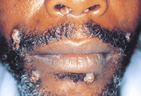
A previously healthy 39-year-old black man presented with a 4-month history of multiple papular and nodular lesions on his scalp, face, and neck, as well as painful lesions in his mouth and on his tongue. About a month before this eruption occurred, the patient experienced swelling of the right side of his neck; the swelling was associated with headache, sore throat, earache, arthralgia, sweats, and temperatures ranging from 38°C to 39°C, all of which lasted around 3 weeks. Several weeks before these symptoms developed, the patient had noticed a painless penile lesion.
Examination revealed multiple flesh-colored to red-brown papules and nodules located on the face, neck, scalp, and nares (Figure 1). Several nodules had a crusted surface. White patches were noticed on the palate, tongue, and lips. There were collarettes of scale on both palms. Bilateral, tender adenopathy was found in the cervical, axillary, epitrochlear, and inguinal regions. Findings of the physical examination were otherwise unremarkable.
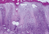
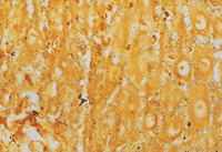
A biopsy specimen was obtained for routine hematoxylin-eosin stain (Figure 2) and special stains (Figure 3).
What is your diagnosis?
From Fort Riley, Kan (Dr Harden), and Fort Sam Houston, Tex (Dr Keeling).
|
Diagnosis and Discussion: Secondary Syphilis
HISTOPATHOLOGIC FINDINGS AND LABORATORY DATA
A dense infiltrate containing many plasma cells was noted in the reticular dermis. A psoriasiform hyperplasia was present, and the epidermis was infiltrated by numerous neutrophils. Some evidence of endothelial swelling of blood vessels was noted. A Dieterle stain demonstrated multiple spirochetes within the epidermis and a few scattered spirochetes within the superficial dermis (Figure 3).
A rapid plasma reagin test was positive for syphilis at a titer of 1:128, and the results of a florescent treponema antibody test were positive. Serologic tests were negative for mononucleosis, antinuclear antibody, and human immunodeficiency virus. A complete blood cell count and chest x-ray film revealed no abnormalities.
DISCUSSION
Nodular lesions of syphilis seem to be uncommon but are periodically reported in the literature.1-9 They may appear on any area of the body, with the head and neck often involved, and generally are red-brown or violaceous. Although these nodules are said to be more common in tertiary syphilis, most recent reports have diagnosed them as a manifestation of secondary syphilis in patients who may also exhibit mild lymphadenopathy without associated constitutional symptoms. The histologic pattern varied, but dermal infiltration of plasma cells, lymphocytes, histiocytes, eosinophils, and/or neutrophils was seen in the majority of cases. Granulomas were seen in some cases.3, 8
Red-brown, vegetative, nodular lesions of syphilis that have an irregular surface have been referred to as framboesiform or raspberrylike.1-2,7 This raspberrylike surface also has been described as being warty, and may also be moist, crusted, or ulcerated. We believe that our patient's nodular lesions could fit the description embodied in this older term, framboesiform syphilis.
The differential diagnosis of nodular secondary syphilis includes deep fungal infection, Kaposi sarcoma, bacillary angiomatosis, foreign body granuloma, lymphoma, lymphomatoid papulosis, pseudolymphoma, leprosy, sarcoidosis, and halogenoderma.
The histologic pattern of secondary syphilis is variable and can mimic many other diseases, such as persistent gyrate erythema, lichen planus, psoriasis, pustular psoriasis, parapsoriasis lichenoides et varioliformis acuta, histiocytoma, and sarcoidosis.10 Common findings include superficial and/or deep perivascular or diffuse dermal infiltrates of lymphocytes, histiocytes, and plasma cells; epidermal hyperplasia with spongioform, psoriasiform, or lichen planus–like appearance; and dilated blood vessels with a swollen endothelial lining.10 Granulomatous infiltrates are sometimes evident, particularly in older lesions.5, 11
With the advent of penicillin and effective serologic screening measures, the incidence of syphilis declined, and until 1985 relatively few cases were diagnosed. Between 1985 and 1990, there was a 75% increase in the incidence of primary and secondary syphilis.12 Syphilis is again becoming a major health problem, and it is important that physicians be aware of the various cutaneous manifestations of this disease for proper diagnosis.
AUTHOR INFORMATION
Selected from Arch Dermatol. 1997;1333:1027. Off-Center Fold.
REFERENCES
|
1. Lejman K, Starzycki Z. Keratopustular variety of framboesiform syphilis: a case report. Br J Venereol Dis. 1977;53:195-199.
2. Beck MH, Hubbard HC, Dave VK, Haye KR. Secondary syphilis with framboesiform facial lesions: a case report. Br J Venereol Dis. 1981;56:103-105.
3. Graham WR Jr, Duvic M. Nodular secondary syphilis. Arch Dermatol. 1982;118:205-206.
FREE FULL TEXT
4. Baum EW, Bernhardt M, Sams WM Jr, Alexander WJ, McLean GL. Secondary syphilis: still the great imitator. JAMA. 1983;249:3069-3070.
FREE FULL TEXT
5. Matsuda-John SS, McElgunn PSJ, Ellis CN. Nodular late syphilis. J Am Acad Dermatol. 1983;9:269-272.
FULL TEXT
|
ISI
| PUBMED
6. Hodak E, David M, Rothem A, Bialowance M, Sandback M. Nodular secondary syphilis mimicking cutaneous lymphorecticular process. J Am Acad Dermatol. 1987;17(pt 2):914-917.
7. Tham SN, Ng SK. Secondary syphilis with framboesiform lesions. Genitourin Med. 1990;66:99-100.
ISI
| PUBMED
8. Pavithran K. Nodular secondary syphilis. Int J Dermatol. 1991;30:799-800.
FULL TEXT
|
ISI
| PUBMED
9. Adriaans B. An erythematous nodular eruption: secondary syphilis. Arch Dermatol. 1992;128:978-981.
FULL TEXT
| PUBMED
10. Jeerapaet P, Ackerman AB. Histologic patterns of secondary syphilis. Arch Dermatol. 1973;107:373-377.
FREE FULL TEXT
11. Abell E, Marks R, Jones EW. Secondary syphilis: a clinico-pathological review. Br J Dermatol. 1975;93:53-61.
ISI
| PUBMED
12. Hook EW, Marra CM. Acquired syphilis in adults. N Engl J Med. 1992;326:1060-1069.
ISI
| PUBMED
|