|
|
Chondrodermatitis Nodularis Chronica Helicis
Thomas J. Zuber, MD;
Edward Jackson, MD
Arch Fam Med. 1999;8:445-447.
ABSTRACT
Chondrodermatitis nodularis chronica helicis is a painful nodule of the external ear. These uncommon lesions are most often encountered on the helix in white men older than 40 years, although they also rarely occur on the antihelix in women. The lesions frequently present with exquisite tenderness that interferes with sleep. While the cause of this dermal inflammatory process is not known, long-term trauma or sun damage may play a role. Recurrences often complicate treatment if all sites of inflammation are not eradicated. Surgical treatment is generally recommended, either by wide excision or by deep shave and treatment of the underlying cartilage.
INTRODUCTION
Chondrodermatitis nodularis chronica helicis (CNCH) describes an uncommon, painful, inflammatory nodule of the external ear.1-8 Most cases involve middle-aged or elderly white men who have these nodules on the outer rim of the helix.1-4,8 Women and nonwhites have been noted occasionally to have lesions in areas other than the helix, such as the antihelix or antitragus. The lesions are believed by several researchers1-3,9 to relate to trauma or sun damage. The nodules are more commonly reported on the right ear, which is believed to be the preferred resting side during sleep.9 The lack of a thick, cushioning, subcutaneous layer in the auricle may predispose the underlying cartilage to pressure-induced ischemia and damage.8 Despite numerous theories, the exact cause and pathogenesis of CNCH remain uncertain.2, 4-5
CLINICAL APPEARANCE
The nodules of CNCH usually appear dome shaped, firm, reddish gray, with an erythematous rim, and vary from 3 to 10 mm (Figure 1). 1-2 The surface of the lesion often is covered with a scale or crust over an underlying central depression.1-2 Ulceration can be present, and the lesion usually is tender to even the slightest pressure. The lesions usually persist, with spontaneous remission rare.1 The lesions do not develop malignancy.
|
|
|
|
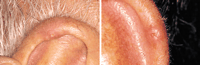
Figure 1. Nodules of chondrodermatitis nodularis chronica helicis on the helix. The lesions appear raised and reddish gray, with a superficial scaling.
|
|
|
Lesions often appear to arise spontaneously, and patients may not relate a definite history of trauma. The nodules often cause notable distress, and patients often seek medical advice when the pain interferes with sleep.9-10 The characteristic location, appearance, and pain usually allow for proper diagnosis.1, 9 Histological examination may support the diagnosis, but is performed mainly to exclude cancer.9
Histologically, the lesions often demonstrate dermal inflammation and fibrosis associated with either a central hyperkeratotic plug or ulceration and crust.1, 3 Ulcer margins often demonstrate hyperplasia, but Hurwitz3 noted that one third of 84 lesions had no ulceration. Cartilage beneath the granulomatous and fibrotic dermis often is disrupted, hemorrhagic, and even necrotic, although occasionally it can appear undamaged.3 Chondrodermatitis nodularis chronica helicis lesions could begin as a perichondritis, extending outward to involve the skin.6 It is possible that inflamed hair follicles in the skin may be central to the pathologic process.3
The 2 conditions most frequently encountered with similar clinical appearance are basal cell and squamous cell carcinoma.1 Squamous cell carcinoma is more common on the auricle,2 and continues in a slow growth pattern, while CNCH often stops growing on reaching its mature size.11 Cancer can be easily differentiated from CNCH by the differing histological appearance on the biopsy specimen. Other lesions in the differential diagnosis include actinic keratoses, cutaneous horns, keratoacanthomas, warts, and elastotic nodules on the antihelix.1, 12 Keratoacanthomas, warts, and cutaneous horns often continue to grow over time, and generally lack notable tenderness.11
MANAGEMENT OPTIONS
The unusual nature of CNCH and the high recurrence rate for this benign condition has produced a wide variety of surgical and nonsurgical treatment options.2, 6 Nonsurgical options include cryotherapy, topical corticosteroids, topical antibiotic ointments, intralesional collagen injection, and intralesional corticosteroid injection.1-5,8, 13 Surgical options are generally considered the treatment of choice, and include wedge resection of the skin and cartilage with suture closure; curettage and electrocautery; carbon dioxide laser ablation therapy; and excision of the skin lesion followed by scalpel resection, curettage, or electrosurgical treatment of the underlying cartilage.1-10,14-16
Historically, topical antibiotic ointments were applied to CNCH to cushion the skin and help reduce trauma to the lesion. Few controlled series have examined any of the topical therapies, including corticosteroids; many experienced researchers1-2,5, 9-10 believe that topical therapy is not a major treatment consideration. Beck13 reported the resolution of CNCH in a small series of 5 patients treated with topical 0.1% betamethasone valerate and 3% clioquinol proprietary cream applied twice daily. Three series from before 1960 described fewer than 20 total patients treated with topical corticosteroids.6
Similarly, only small studies have examined injection therapy for CNCH. Greenbaum4 reported complete symptom relief in 5 patients following injection of collagen implants, suggesting possible benefit produced by improved cushioning of the underlying cartilage. Corticosteroid injections have been reported to produce some benefit, usually using the injection of 0.1 to 0.2 mL of triamcinolone acetonide (10-40 mg/mL).1, 5, 16 Lawrence16 noted symptom resolution in only 12 (27%) of 44 injected ears with CNCH. While many patients who receive corticosteroid injections may eventually require excisional therapy, this nonsurgical intervention is advocated by some as an initial treatment option.1
Physicians may need to perform a biopsy of lesions of CNCH to exclude the presence of other conditions such as cancer. Shave biopsy followed by treatment to the wound base is often advocated as an initial diagnostic and therapeutic intervention.2, 9 Soft, necrotic, underlying cartilage can be removed by vigorous curettage.2 Reports from before 1960 suggested recurrences from this technique at more than 20%.6, 9 More aggressive treatment to the underlying cartilage also has been performed. Kromann et al9 reported a recurrence rate of 31% among 142 patients treated with curettage followed by electrodesiccation (electrosurgical ablation) of the underlying cartilage. We have successfully treated 4 patients with a modification of the shave technique on the skin, followed by electrosurgical feathering of the base (Figure 2). Electrosurgical feathering allows for treatment of partial thickness of the cartilage, and treatment of surrounding skin. To our knowledge, no large series have been published on this technique.
|
|
|
|
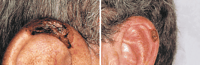
Figure 2. The skin nodules have been removed by the shave technique, the cartilage shaved, and electrosurgical feathering performed to remove any residual diseased tissue. The edges of the defect have been smoothed (feathered) to produce a cosmetically acceptable final appearance.
|
|
|
Surgical excision remains the hallmark of therapy for CNCH.1 Unfortunately, recurrences are common if all sites of inflammation are not removed.2, 6, 10 Even when wide excision of the underlying cartilage is performed, recurrences have developed in up to 10% of patients at the edges of the excised cartilage.6, 15 The cartilage damage beneath a nodule usually involves only 1 cartilage surface, and some physicians advocate a partial excision procedure involving the cartilage, rather than a through-and-through pie-shaped or wedge excision of the auricle.
A variation of the shave technique for removal of the nodule of CNCH involves fusiform excision of the skin nodule, and then horizontal shaving of the underlying cartilage.8, 10, 16 By undermining the skin and perichondrium off the underlying cartilage for 1 cm on both sides of the skin excision, the skin flaps allow for good visualization of the cartilage for appropriate trimming (Figure 3).6 The skin edges are then closed primarily with this technique. Lawrence16 has advocated that only the damaged cartilage should be removed, with skin flap closure performed over the excised cartilage. Lawrence has reported excellent cosmetic results and cures in 34 (74%) of 46 patients.
|
|
|
|
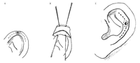
Figure 3. A, Planned excision lines along the helix. B, The lesion is excised, the skin flaps created, the cartilage treated, and skin closed. C, planned excision lines on the antihelix. Adapted from the technique described by Munnoch et al.6
|
|
|
SUMMARY
Chondrodermatitis nodularis chronica helicis is an uncommon dermatologic condition that can present to the family physician as a painful nodule on the ear. Proper identification of the lesion, and exclusion of a skin cancer of the auricle, will allow for proper treatment. Family physicians may initially try topical or injection therapy, but surgical therapy usually is required. Several fairly simple excisional procedures can be performed to remove the painful nodule, but additional treatment of the underlying diseased cartilage will provide the highest cure rates.
AUTHOR INFORMATION
Accepted for publication February 4, 1999.
We thank Stephanie John, MLn, DM, AHIP, and the Medical Informatics Team at Saginaw Cooperative Hospitals, Saginaw, Mich, for research assistance.
Corresponding author: Thomas J. Zuber, MD, Saginaw Cooperative Hospitals, 1000 Houghton Ave, Saginaw, MI 48602 (e-mail: zuber{at}pilot.msu.edu).
From the Department of Family Medicine and the Family Practice Residency Program, Saginaw Cooperative Hospitals (Drs Zuber and Jackson), and the Dermatology Clinic, Aleda E. Lutz Veterans Affairs Medical Center (Dr Zuber), Saginaw, Mich; and Department of Family Medicine, Michigan State University, Lansing (Drs Zuber and Jackson).
REFERENCES
| |
1. Wade TR. Chondrodermatitis nodularis chronica helicis: a review with emphasis on steroid therapy. Cutis. 1979;24:406-409.
PUBMED
2. Habif TP. Clinical Dermatology: A Color Guide to Diagnosis and Therapy. 3rd ed. St Louis, Mo: Mosby–Year Book Inc; 1996:643.
3. Hurwitz RM. Painful papule of the ear: a follicular disorder. J Dermatol Surg Oncol. 1987;13:270-274.
PUBMED
4. Greenbaum SS. The treatment of chondrodermatitis nodularis chronica helicis with injectable collagen. Int J Dermatol. 1991;30:291-294.
PUBMED
5. Coldiron BM. The surgical management of chondrodermatitis nodularis chronica helicis. J Dermatol Surg Oncol. 1991;17:902-904.
PUBMED
6. Munnoch DA, Herbert KJ, Morris AM. Chondrodermatitis nodularis chronica helicis et antihelicis. Br J Plast Surg. 1996;49:473-476.
PUBMED
7. Taylor MB. Chondrodermatitis nodularis chronica helicis successful treatment with the carbon dioxide laser. J Dermatol Surg Oncol. 1991;17:862-864.
PUBMED
8. Long D, Maloney ME. Surgical pearl: surgical planing in the treatment of chondrodermatitis nodularis chronica helicis of the antihelix. J Am Acad Dermatol. 1996;18:761-762.
9. Kromann N, Hoyer H, Reymann F. Chondrodermatitis nodularis chronica helicis treated with curettage and electrocauterization: follow-up of a 15-year material. Acta Derm (Stockh). 1983;63:85-87.
10. Sinclair P. Excision technique for chondrodermatitis nodularis helicis. Australas J Dermatol. 1996;37:61.
PUBMED
11. Beilan B. What's your assessment? Dermatol Nurs. 1997;9:239, 282.
PUBMED
12. Weedon D. Elastotic nodules of the ear. J Cutan Pathol. 1981;8:429-433.
FULL TEXT
| PUBMED
13. Beck MH. Treatment of chondrodermatitis nodularis helicis and conventional wisdom? Br J Dermatol. 1985;113:504-505.
PUBMED
14. Karam F, Bauman T. Carbon dioxide laser treatment for chondrodermatitis nodularis chronica helicis. Ear Nose Throat J. 1988;67:757-763.
PUBMED
15. Kitchens GG. Auricular wedge resection and reconstruction. Ear Nose Throat J. 1989;68:673-683.
PUBMED
16. Lawrence CM. The treatment of chondrodermatitis nodularis with cartilage removal alone. Arch Dermatol. 1991;127:530-535.
FREE FULL TEXT
RELATED ARTICLE
The Archives of Family Medicine Continuing Medical Education Program
Arch Fam Med. 1999;8(5):383-385.
FULL TEXT
THIS ARTICLE HAS BEEN CITED BY OTHER ARTICLES
Answer: Can you identify this condition?
Devani and Barankin
cfp 2007;53:837-837.
FULL TEXT
|