|
|
An Ulcerated Nodule Associated With Lymphadenopathy
Sarah Boyce, BS;
José R. Peña, MD;
Daniel A. Davis, MD
Arch Fam Med. 2000;9:316-317.
REPORT OF A CASE
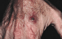
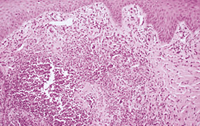
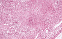
A 37-year-old male patient who had undergone renal transplantation presented with a 1-week history of a persistent fever (temperatures to 39°C). Physical examination revealed an ulcerated nodule over the right second metacarpal bone (Figure 1) and a 5-cm, firm, tender lymph node in the right axilla. The nodule developed in a scratch the patient had gotten while working in his rose garden. His adult cat had subsequently licked the wound. Excisional biopsy specimens were obtained from the cutaneous nodule (Figure 2) and the lymph node (Figure 3). The lymph node specimen was also stained with the Warthin-Starry silver preparation.
What is your diagnosis?
From the University of Alabama, Birmingham.
Diagnosis and Discussion: Cat-Scratch Disease
HISTOPATHOLOGIC FINDINGS, LABORATORY DATA, AND CLINICAL COURSE
Routine histologic examination of the cutaneous nodule showed dermal abscesses surrounded by a halo of macrophages. Stains for infectious organisms (Fite, Gomori methenamine-silver, Warthin-Starry, and Gram) were noncontributory. Routine histologic examination of the axillary lymph node showed expanded paracortical areas with focal necrosis and a mixed infiltrate. A Warthin-Starry preparation stained clusters of bacilli within the necrotic foci.
The patient's leukocyte count was normal, as was his differential cell count. No bacteria, fungi, or mycobacteria were cultured from blood or lymph node tissue. No antibodies to toxoplasma, histoplasma, blastomyces, cryptococcus, human immunodeficiency virus, or cytomegalovirus were found. IgG antibodies (enzyme-linked immunosorbent assay) to Bartonella were present at a titer of 1:77 (reference range, <1:12).
The patient's fever rapidly resolved with oral erythromycin therapy. At 3 months, his disease appeared clinically resolved. Follow-up Bartonella titers are not available.
DISCUSSION
In 1889, Parinaud1 first reported an oculoglandular syndrome of lymphadenopathy and conjunctivitis that is now known as cat-scratch disease (CSD). In 1950, Debré et al2 described the relationship of adenopathy and house-cat contact.
Several genetically related organisms have been implicated in the etiology of CSD. Initially, Afipia felis was considered the causative agent. However, patients with CSD did not mount antibody responses, determined by antibody titer, against A felis. Subsequently, Rochalimaea species, gram-negative bacillary  -proteobacteria originally named after Rocha-Lima, were isolated from and associated with an immune response in patients with CSD.3 It should be mentioned that Rochalimaea species have been renamed based on extensive 16S rRNA sequence homology with organisms in the genus Bartonella.4 Thus, it now appears that CSD is caused almost exclusively by Bartonella henselae, named after Alberto Barton and Diane Hensel.5 -proteobacteria originally named after Rocha-Lima, were isolated from and associated with an immune response in patients with CSD.3 It should be mentioned that Rochalimaea species have been renamed based on extensive 16S rRNA sequence homology with organisms in the genus Bartonella.4 Thus, it now appears that CSD is caused almost exclusively by Bartonella henselae, named after Alberto Barton and Diane Hensel.5
The cutaneous lesion of CSD is typically a nodule that develops 1 week after traumatic contact with a cat.3, 6 Despite an earlier designation as cat-scratch fever, malaise, headache, and fever occur in fewer than 50% of patients.6-7 Lymphadenopathy, typically axillary, is essentially the sine qua non of CSD and occurs within 3 weeks of inoculation. Eighty-five percent of patients have only a single involved node.7 The course of CSD is usually benign and often self-resolving, but complications such as encephalitis, osteomyelitis, and hepatosplenomegaly have been described in as many as 20% of patients.3, 7
In the past, histopathologic examination of the involved lymph node specimens was thought to be the most reliable diagnostic test for CSD. Key features include stellate caseating granulomas, microabcesses, and lymphoid follicular hyperplasia.8 Only weakly gram negative, Bartonella organisms routinely stain with the Warthin-Starry silver preparation. Although the organisms are technically culturable from specimens of tissue and blood, either typical hospital laboratories do not use appropriate media to isolate B henselae or they discard cultures sooner than is required for isolation.3 However, other diagnostic methods, such as the polymerase chain reaction, as well as the indirect fluorescent antibody technique and the enzyme-linked immunosorbent assay, both of which can measure serum antibody levels, have recently been developed.3, 6, 9 It has been suggested that the indirect fluorescent antibody technique is presently the most effective test, with one study showing up to 93% sensitivity and 98% specificity in selected populations.9
Cat-scratch disease is most often seen in children or in association with human immunodeficiency virus infection.5 Bartonella infection associated with other forms of immunosuppression is exceedingly rare.10 This case is also unusual in that trauma and inoculation were sequential and not simultaneous.
Sporotrichosis is the other major consideration in the clinical differential diagnosis. Sporothrix can be inoculated by rose thorn puncture, and the domestic cat is a known sporothrix vector.11
AUTHOR INFORMATION
Selected from Arch Dermatol. 1999;135:983. Off-Center Fold.
REFERENCES
|
1. Parinaud H. Conjonctivité infectieuse transmise par les animaux. Ann Oculistique. 1889;101:252-253.
2. Debré R, Lamy M, Jammet M, Costil L, Mozzconacci P. La maladie des griffes de chat. Semaine Hopitaux Paris. 1950;26:1895-901.
3. Jerris RC, Regnery RL. Will the real agent of cat-scratch disease please stand up? Annu Rev Microbiol. 1996;50:707-725.
FULL TEXT
|
ISI
| PUBMED
4. Brenner DJ, O'Connor SP, Winkler HH, Steigerwalt AG. Proposals to unify the genera Bartonella and vinsonii comb. nov., Bartonella henselae comb. nov., and Bartonella elizabethae comb. nov., and to remove the family Bartonellaceae from the order Rickettsiales. Int J Syst Bacteriol. 1993;43:777-786.
FREE FULL TEXT
5. Regnery RL, Anderson BE, Clarridge III JE, Rodriguez-Barradas MC, Jones DC, Carr JH. Characterization of a novel Rochalimaea species, R. henselae, sp. nov., isolated from blood of a febrile, human immunodeficiency virus–positive patient. J Clin Microbiol. 1992;30:267-274.
6. Jevon G, Dunne WM, Finegold MJ. An analysis of lymph node DNA for possible bacterial agents of cat-scratch disease. Pediatr Pathol Lab Med. 1995;15:3-9.
ISI
| PUBMED
7. Carithers HA, Carithers CM, Edwards RO. Cat-scratch disease: its natural history. JAMA. 1969;207:312-316.
FREE FULL TEXT
8. Adal KA, Cockerell CJ, Petri WA Jr. Cat-scratch disease, bacillary angiomatosis, and other infections due to Rochalimaea. N Engl J Med. 1994;330:1509-1515.
FREE FULL TEXT
9. Szelc-Kelly CM, Goral S, Perez-Perez GI, Perkins BA, Regnery RL, Edwards KM. Serologic responses to Bartonella and Afipia antigens in patients with cat scratch disease. Pediatrics. 1995;96:1137-1142.
FREE FULL TEXT
10. Torok L, Viragh S, Borka I, Tapai M. Bacillary angiomatosis in a patient with lymphocytic leukaemia. Br J Dermatol. 1994; 130:665-668.
11. Dunstan RW, Langham RF, Reimann KA, Wakenell PS. Feline sporotrichosis: a report of five cases with transmission to humans. J Am Acad Dermatol. 1986;15:37-45.
ISI
| PUBMED
|