|
|
Infantile Henoch-Schönlein Purpura
Avinash K. Shetty, MD;
Bonnie C. Desselle, MD;
John L. Ey, MD, MPH;
Hernan Correa, MD;
Wesley K. Galen, MD;
Abraham Gedalia, MD
Arch Fam Med. 2000;9:553-556.
ABSTRACT
Henoch-Schönlein purpura is a common cause of vasculitis in children. This condition is unusual in infants and children younger than 2 years. We describe a 4-month-old infant with infantile Henoch-Schönlein purpura and review the clinical spectrum, differential diagnoses, and the histopathologic features of the disease. Its relations to Henoch-Schönlein purpura in older children are discussed.
INTRODUCTION
Henoch-Schönlein purpura (HSP) is one of the most common vasculittides of childhood, characterized by the classic triad of nonthrombocytopenic purpura, colicky abdominal pain, and arthritis. The syndrome of acute purpura and arthritis in children was first described by Schönlein in 1837 and the manifestations of abdominal pain and nephritis were added by Henoch in 1874.1 It occurs most often in children between the ages of 3 and 10 years. The occurrence of HSP in infants or young children is rare. This entity was first described in infants in the United States by Snow in 1913.2 Subsequently, most cases have appeared in the Eurpoean literature. We describe herein a 4-month-old infant with infantile HSP who presented with fever and purpuric rash. We review the clinical manifestations, differential diagnoses, and histopathological features of this disease in infants and older children.
REPORT OF A CASE
A 4-month-old African American female infant was admitted with a 1-day history of fever, increased irritability, decreased appetite, and purpuric rash. Several small papular lesions initially appeared on the lower extremities. These lesions enlarged, changed from red to purple, and spread to the upper extremities, face, and ears. She was the product of a normal-term gestation. There was no other significant medical history.
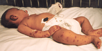
On physical examination, she was alert, well hydrated, and afebrile. A diffuse, symmetric palpable purpuric rash involving the face, ears, and extremities was visible (Figure 1). There were large ecchymoses on the left helix and fingers of both hands and toes. No subcutaneous edema was noted. The rest of the physical examination yielded normal findings. Results of the initial laboratory studies included hemoglobin level of 110 g/L, white blood cell count of 14.3 x 10⁹/L (0.39 segmented neutrophils, 0.10 band forms, 0.35 lymphocytes, 0.15 monocytes, and 0.01 eosinophils), and a platelet count of 842 x109/L. Prothrombin time, partial thromboplastin time, levels of serum electrolytes and serum urea nitrogen, and results of urinalysis, stool guaiac test, and cerebrospinal fluid analysis were normal.
|
|
|
|
Figure 1. Palpable purpuric rash on face, ears, and extremities in a 4-month-old infant.
|
|
|
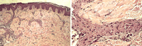
The patient was admitted to the pediatric intensive care unit with a presumptive diagnosis of meningococcemia, and intravenous ceftriaxone sodium (100 mg/kg per day) was administered empirically. The initial blood, urine, and cerebrospinal fluid cultures were negative for pathogens. The patient clinically looked well, with no obvious systemic symptoms. At this point, a punch biopsy specimen of the skin lesion was obtained; histopathologic findings are shown in Figure 2. Staining of sections with hematoxylin-eosin revealed perivascular fragmentation of inflammatory cell nuclei and intraluminal fibrin thrombi affecting small blood vessels, including the postcapillary venules in the upper and middle dermis, a classic sign of leukocytoclastic vasculitis.3 There was evidence of polymorphonuclear leukocyte infiltration throughout the vessel wall. Results of IgA immunofluorescence were negative. In view of the patient's presentation, clinical course, culture findings, and leukocytoclastic vasculitis, a diagnosis of infantile HSP was made, and the patient was discharged home after 10 days of hospitalization. At follow-up 3 weeks later, the rash had resolved, and the infant was well with no sequelae.
|
|
|
|
Figure 2. Left, Biopsy specimen of skin lesion demonstrates perivascular leukocytoclastic changes and intraluminal thrombi (hematoxylin-eosin, original magnification x100). Right, Dermal vessel with perivascular leukocytoclastic changes and intraluminal thrombi (hematoxylin-eosin, original magnification x400).
|
|
|
COMMENT
Henoch-Schönlein purpura is the most common systemic vasculitic disease of childhood involving small vessels, especially the skin.4-5 It has an annual incidence of approximately 14 cases per 100,000 population, is more common in boys, and peaks during the winter.4 Henoch-Schönlein purpura occurs most often in children aged 3 through 10 years and is characterized by the classic triad of nonthrombocytopenic palpable purpuric rash, abdominal pain or renal involvement, and arthritis.4-5 The etiology of HSP is unknown, although there seems to be a temporal relationship with recent respiratory infections. Group A streptococcal infections have been implicated, but other organisms including adenovirus, parvovirus B19, and mycoplasma have also been reported to precede HSP.5 Exposure to allergens in medications (penicillin, sulfonamides, allopurinol, propylthiouracil, and quinidine) or food, exposure to cold, and insect bites have been implicated in some cases.4, 6-7 Most cases occur in the winter and spring months, supporting an infectious trigger in a susceptible individual. The diagnosis of HSP is made usually after the appearance of the classic palpable purpuric rash that primarily affects the lower extremities and buttocks. Gastrointestinal tract disease occurs in approximately two thirds of these children, varying from colicky abdominal pain, nausea, and vomiting to intestinal hemorrhage, intussusception, pancreatitis, and hydrops of the gallbladder.5, 8 Abdominal symptoms may precede the typical purpuric rash of HSP in 14% to 36%, making initial diagnosis difficult and even resulting in unnecessary laparotomy.8 Joint involvement, which occurs in 60% to 84% of cases, is transient, generally affecting the ankles and knees, and leaves no permanent deformity.5 The most serious complication of HSP is renal involvement, which occurs in 50% of older children but is only serious in some 10% of patients. In 80% of those with renal involvement, it becomes apparent within the first 4 weeks of illness.5 The spectrum of renal disease varies from isolated microscopic hematuria to a nephritis or nephrotic syndrome with renal failure. Overall, 2% to 5% of cases progress to end-stage renal failure.9 Scrotal involvement is not uncommon and may mimic testicular torsion, which must be excluded. Other unusual complications of HSP include florid cerebral manifestations, including seizures, paresis, or coma; cholecystitis; myocardial infarction; and interstitial lung disease.5 Treatment is supportive, as this is a self-limiting disease; corticosteroids may be beneficial in patients with severe abdominal pain, but are not recommended for treatment of rash, joint pain, or renal disease alone.4 The disease may last for 3 to 6 weeks, with recurrence in up to 50% of patients.
The occurrence of HSP in infants or young children is rare.10-12 Although first described by Snow2 in 1913 and subsequently in frequent reports in the European literature under a variety of headings (ie, acute hemorrhagic edema of infancy [AHEI],13 Finkelstein disease,14 Seidelmayer syndrome,15 and infantile postinfectious iris–like purpura and edema16), the entity is not well recognized in the English-language literature.17 The clinical spectrum of HSP in younger children differs from that in older children (Table 1).18 It typically affects infants aged 4 to 24 months, often after an acute respiratory tract infection and/or drug ingestion, with a dramatic onset of acute palpable purpura, ecchymoses, and tender edema of the limbs and face.10-11,19 Fever, if present, is usually low, and the patients are hemodynamically stable.10 The principal cutaneous lesions often have a cockade (medallionlike) rosette-shaped pattern on the face, auricles, and extremities. The lesions usually appear in successive crops and display variable stages of evolution at any given time.17 This eruption is clinically distinct from the palpable purpuric lesions on the extensor surfaces of the extremities and buttocks in older children with HSP.17 Subcutaneous edema is more common in infants than in children older than 2 years.18 Urticaria, petechiae, and ear lobe necrosis constitute additional rare skin manifestations of infantile HSP.12 Unlike older children in whom visceral involvement (gastrointestinal tract bleeding, arthritis, and nephritis) is common, in infantile HSP it is unusual.17
|
|
|
|
Table 1. Clinical Features of Henoch-Schönlein Purpura
|
|
|
There is disagreement to whether AHEI is part of the spectrum of HSP or a separate disorder. The younger age of patients with AHEI; the extensive edema with face, ear, and limb purpura; rare visceral involvement; and infrequent relapses have led some authors to suggest that AHEI may be a distinct clinicopathologic entity and not a variant of HSP.10, 20 On the other hand, some investigators believe that the distinct features of HSP in infants are partially explicable by age and provide another example of the wide clinical spectrum of a disease entity.2, 11-12 The reason for the frequency of facial purpura in infants with HSP is unclear. Amitai et al12 have postulated that the proportionally larger head and face with a corresponding increase in blood supply in infants would render them more susceptible to facial purpura.
The combination of fever and a purpuric skin rash constitutes a diagnostic challenge for the clinician (Table 2). Serious and potentially life-threatening purpuric disorders, particularly sepsis resulting from meningococcemia, endocarditis, Rocky Mountain spotted fever, as well as other vasculitic syndromes such as systemic lupus erythematosus, clotting disorders, or thrombocytopenia must be excluded promptly.4-5 The clinical findings, particularly the distribution of the rash, with results of hematologic studies should identify these patients.5 The diagnosis of HSP in children requires that only palpable purpura with a normal platelet count be documented.21-22 There are no specific diagnostic laboratory markers for HSP. Erythrocyte sedimentation rates are usually normal or minimally elevated. Antinuclear antibody and rheumatoid factor are not present. Serum IgA levels are increased in about 50% of patients during the acute illness. In atypical cases in which the diagnosis is in doubt, a biopsy of the skin rash may be necessary. Histopathologic features of the skin lesions in infantile HSP can range from a typical leukocytoclastic vasculitis with or without fibrinoid necrosis to the less specific findings of a lymphohistiocytic perivascular infiltrate with extravasation of erythrocytes.17 The pathogenic lesions seen in HSP are probably immune mediated. In older children with HSP, immunofluorescence studies demonstrate perivascular IgA deposition in almost 100% of cases,17 whereas this finding is rare in infantile HSP, although C3 and IgM are more commonly found in the affected vessel walls.10-11 Circulating IgA immune complexes may be present in some patients,11 although data supporting the presence of classic antigen-antibody complexes have been questioned. Possibly, IgA aggregates or IgA complexes with complement are being detected in target organs, resulting in elaboration of inflammatory mediators, including vascular prostaglandins such as prostacyclin, that may play a central role in the pathogenesis of vasculitis.23
|
|
|
|
Table 2. Differential Diagnosis of Fever and Purpuric Rash
|
|
|
Infantile HSP is a self-limited disease of short duration, with spontaneous remission usually expected in 1 to 3 weeks.17 However, recurrence is possible.11 There is no effective therapy, and systemic steroids do not seem to alter the course of the disease.12
AUTHOR INFORMATION
Accepted for publication January 28, 2000.
Corresponding author: Abraham Gedalia, MD, Louisiana State University Medical Center, Department of Pediatrics, 1542 Tulane Ave, T8-1, New Orleans, LA 70112 (e-mail: a61543{at}pol.net).
From the Departments of Pediatrics (Drs Shetty, Desselle, Ey, and Gedalia), Pathology (Dr Correa), and Dermatology (Dr Galen), Louisiana State University Medical Center and Children's Hospital, New Orleans.
REFERENCES
| |
1. Henoch EH. Uber ein eigenthümliche Form von Purpura. Berl Klin Wochenschr. 1874;11:641-643.
2. Snow IM. Purpura, urticaria and angioneurotic edema of the hands and feet in a nursing baby. JAMA. 1913;61:18-19.
FREE FULL TEXT
3. Lie JT. American College of Rheumatology Subcommittee on Classification of Vasculitis: illustrated histopathologic classification criteria for selected vasculitic syndromes. Arthritis Rheum. 1990;33:1074-1087.
ISI
| PUBMED
4. Kraft DM, Mckee D, Scott C. Henoch-Schönlein purpura: a review. Am Fam Physician. 1998;58:405-408.
ISI
| PUBMED
5. Tizard EJ. Henoch-Schönlein purpura. Arch Dis Child. 1999;80:380-383.
FREE FULL TEXT
6. Szer IS. Henoch-Schönlein purpura. Curr Opin Rheumatol. 1994;6:25-31.
PUBMED
7. Ansell BM, Falcini F. Cutaneous vasculitis in children. Clin Dermatol. 1999;17:577-580.
FULL TEXT
|
ISI
| PUBMED
8. Choong CK, Beasley SW. Intra-abdominal mainifestations of Henoch-Schönlein purpura. J Paediatr Child Health. 1998;34:405-409.
FULL TEXT
|
ISI
| PUBMED
9. Koskimies O, Mir S, Rapaola J, Vilska J. Henoch-Schönlein nephritis: long-term prognosis of unselected patients. Arch Dis Child. 1981;56:482-484.
FREE FULL TEXT
10. Saraclar Y, Tinaztepe K, Adalioglu G, Turner A. Acute hemorrhagic edema of infancy (AHEI): a variant of Henoch-Schönlein purpura or a distinct clinical entity? J Allergy Clin Immunol. 1990;86:473-483.
FULL TEXT
|
ISI
| PUBMED
11. Legrain V, Lejean S, Taieb A, et al. Infantile acute hemorrhagic edema of the skin: study of 10 cases. J Am Acad Dermatol. 1991;24:17-22.
ISI
| PUBMED
12. Amitai Y, Gillis D, Wasserman D, Kochman RH. Henoch-Schönlein purpura in infants. Pediatrics. 1993;92:865-867.
FREE FULL TEXT
13. Larregue M, Lesage B, Rossier A. Acute hemorrhagic edema in infants (iris-like purpura with Seidelmayer's post-infectious purpura) and allergic vasculitis [in Spanish]. Med Cutan Ibero Lat Am. 1974;11:165-174.
14. Postma C, Deelman H. Een geval van acuut haemorrhagisch oedeem (Finkelstein). Ned Tijdschr Geneeskd. 1954;98:2592-2595.
15. Leiber B, Olvbrich G. Seidelmayer syndrome. In: Die Klinischen Syndrome. Baltimore, Md: Urban & Schwarzenberg; 1966;1:652.
16. Seidelmayer H. Die fruhinfantle, postinfektrose Kokarden-Purpura. Z Kinderheilkd. 1939;61:217-255.
FULL TEXT
17. Millard T, Harris A, MacDonald D. Acute infantile hemorrhagic oedema. J Am Acad Dermatol. 1999;41:837-839.
FULL TEXT
|
ISI
| PUBMED
18. Allen DM, Diamond LK, Howell DA. Anaphylactoid purpura in children. (Schönlein Henoch syndrome). AJDC. 1960;99:833-854.
19. Dubin BA, Bronson DM, Eng AM. Acute hemorrhagic edema of childhood: an unusual variant of leukocytoclastic vasculitis. J Am Acad Dermatol. 1990;23:347-350.
ISI
| PUBMED
20. Lantner RR, Ros SP. Acute hemorrhagic edema of infancy. Pediatr Emerg Care. 1996;12:111-112.
FULL TEXT
|
ISI
| PUBMED
21. Saulsbury FT. Henoch-Schönlein purpura. Pediatr Dermatol. 1984;1:195-201.
PUBMED
22. Mills JA, Michel BA, Bloch DA, et al. The American College of Rheumatology 1990 criteria for the classification of Henoch-Schönlein purpura. Arthritis Rheum. 1990;33:1114-1121.
ISI
| PUBMED
23. Walker WA, Higuchi LM. Henoch-Schönlein syndrome. In: McMillan JA, ed. Oski's Pediatrics Principles and Practice. 3rd ed. Philadelphia, Pa: Lippincott Williams & Wilkins; 1999:2176-2179.
RELATED ARTICLE
The Archives of Family Medicine Continuing Medical Education Program
Arch Fam Med. 2000;9(6):551-552.
FULL TEXT
THIS ARTICLE HAS BEEN CITED BY OTHER ARTICLES
Postlicensure Safety Surveillance for 7-Valent Pneumococcal Conjugate Vaccine
Wise et al.
JAMA 2004;292:1702-1710.
ABSTRACT
| FULL TEXT
|