


Review of Maxillary Expansion Appliance Activation Methods: Engineering and Clinical Perspectives
- D. L. Romanyk dromanyk{at}ualberta.ca1
- M. O. Lagravere mlagravere{at}ualberta.ca2
- R. W. Toogood roger.toogood{at}ualberta.ca3
- P. W. Major major{at}ualberta.ca2
- J. P. Carey jason.carey{at}ualberta.ca1
- 1Department of Mechanical Engineering, Faculty of Engineering, University of Alberta, 5-8T Mechanical Engineering Building, Edmonton, AB, Canada, T6G 2G8
- 2Orthodontic Graduate Program, Department of Dentistry, Dentistry/Pharmacy Centre, Faculty of Medicine and Dentistry, University of Alberta, Edmonton, AB, Canada, T6G 2N8
- 3Department of Mechanical Engineering, Faculty of Engineering, University of Alberta, 4-8H Mechanical Engineering Building, Edmonton, AB, Canada, T6G 2G8
Abstract
Objective. Review the reported activation methods of maxillary expansion devices for midpalatal suture separation from an engineering perspective and suggest areas of improvement. Materials and Methods. A literature search of Scopus and PubMed was used to determine current expansion methods. A U.S. and Canadian patent database search was also conducted using patent classification and keywords. Any paper presenting a new method of expansion was included. Results. Expansion methods in use, or patented, can be classified as either a screw- or spring-type, magnetic, or shape memory alloy expansion appliance. Conclusions. Each activation method presented unique advantages and disadvantages from both clinical and engineering perspectives. Areas for improvement still remain and are identified in the paper.
1. Introduction
The first recorded maxillary expander, as developed by Angell in 1860, consisted of a shaft with tubular nuts that was rotated using a wrench made from a dime [1]. Many orthodontists have followed suit in using various types of appliances and designs to widen a narrow maxilla. While there are a variety of methods for anchoring the expansion devices, the following paper will only be concerned with activation methods [2, 3]; furthermore, protocols of use of the appliance are not considered in the discussion.
To date, all review publications on the subject are concerned with the clinical impact of expansion while none has reviewed the appliances from a combined clinical and engineering perspective [4, 5]. The purpose of this paper is to analyze current maxillary expansion devices using engineering principles in describing the clinical implications of the activation method while suggesting future areas of design improvement. Only devices concerned with separating the midpalatal suture are considered in the discussion.
2. Materials and Methods
Scopus and PubMed were used to retrieve the literature regarding maxillary expansion appliances. Keywords used in the databases are “maxillary expansion” and “palatal expansion”. Further result reduction was attained by adding “appliance”, “apparatus”, or “device” keywords. A sample search could be given as “maxillary expansion” and “device”, thus allowing for a maximum number of relevant papers to be retrieved from the databases.
Canadian and U.S. patent searches were conducted. In the U.S., the patent classification number used was 433/7 which is defined under the heading of Orthodontics as, “By device having means to apply outwardly directed force (e.g., expander).” In Canada, the current International Classification of A61C7/10 was used along with previous Canadian Classification 83–1.
3. Results
Upon reviewing the literature, it was determined that the methods of activation could be broken down into four categories: screw-type, spring-type, magnetic, and Shape Memory Alloy (SMA) activation methods. Table 1 lists the general activation methods discussed in this paper along with specific examples that fit under these general categories.
Categories of activation methods and specific examples.
Screw-type activation includes any method that requires adjustment through manual rotation of a shaft to expand the appliance. The Hyrax screw (jackscrew), or expansion screw, is commonly seen in current appliances such as those presented by Haas [2] or Biederman [6]. Other typical expansion mechanisms included are car-jack style and telescoping appliances [3, 7]. Whether by a key or wrench, these expanders require frequent patient or clinician adjustment to achieve expansion of the maxilla.
Any mechanism that deforms a body and, subsequently, relies upon elastic restoration forces for maxillary expansion, was classified as a spring-type appliance. This would include devices that utilize coil or wires springs, with representative examples being the Minne Expander [8] and the appliance presented by Defraia et al. [9], respectively. Upon activation, these devices will exert a continuous, yet displacement-dependent, force as the maxilla widens.
Maxillary expansion appliances that utilize magnets as the primary activation method have been reported [10]. Since a magnetic field has directionality, two magnets can be oriented such that they apply opposing forces.
SMA technology is the fourth maxillary expansion activation method. The superelastic nature of these alloys makes them useful in many applications, including orthodontics. In terms of maxillary expansion, it was found that appliances made use of the SMA property through springs—either coil, Darendeliler and Lorenzon [11], or wire, Corbett [12], or expansion screws, Wilchelhaus et al. [13].
4. Discussion
The following will provide further analysis of the four activation methods, while citing specific advantages and disadvantages. Engineering principles related to material and tissue behavior will be used to discuss both the activation method and its clinical impact.
4.1. Midpalatal Suture Behavior
Midpalatal suture structure, and its response to forces, has been discussed in the literature by authors such as Persson [14] and Ten Cate et al. [15]. Also, Bell [16] and Storey [17] have discussed the use of slower expansion using lower forces to obtain more physiologic expansion. As discussed by Persson, studies have shown suture structure to be highly variable at different ages. Additionally, the fiber bundles found inside the suture will change orientation during treatment as a result of the expansion forces applied by the appliance. These complicated structure and behavior greatly affect the mechanics of the maxilla complex during treatment. To the knowledge of the author, there has been no work conducted to obtain a force-displacement or force-time relationship for the unfused suture. Computational models exist that study the mechanical response of the skull to maxillary expansion, but none incorporate the viscoelastic behavior of the sutures [18, 19]. For instance, Lee et al. constructed a finite element analysis model to simulate maxillary expansion that used accepted linear elastic properties of the periodontal ligament for the sutures and not a viscoelastic model [19].
Typical engineering materials, such as steel or aluminum, have linear force-displacement relationships in their elastic range allowing for simple prediction of behavior [20]. Soft tissues exhibit nonlinear viscoelastic force-displacement behavior as shown in Figure 1, which are difficult to predict as they are deformation and deformation rate dependent [21]; the latter produces larger force values at increased deformation rates as illustrated in Figure 1. Since the suture structure is composed of several different soft tissues in a complex geometry, quantitative prediction of force-displacement behavior becomes exceedingly difficult. Furthermore, living tissues are affected by lifestyle choices such as diet and exercise with one example being the effect vitamin intake and exercise level has on bone growth [22].
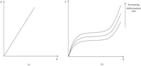
Typical force versus displacement characteristics for a linear (a) and a viscoelastic (b) material.
4.2. Force Application
The behavior of force with respect to displacement will be considered for each of the activation methods. Forces generated in the maxilla by a screw-type appliance, with respect to both time and displacement, are essentially a function of the tissue properties of the patient. The appliance may undergo minor deformation, but it will be negligible compared to that of the maxilla. This is due to the fact that for the same applied force, the ratio of appliance-to-tissue deformation is inversely related to the ratio of their stiffness values, where the stiffness of the appliance will be much greater than that of the soft tissue. Force generation can be visualized by turning an expansion screw in a device that is not placed in a patient. No transverse force is generated as the appliance expands since there is no resistance to expansion other than the friction between screw threads. As such, it is impossible to predict the force that will be generated with respect to time or displacement without fully understanding the properties and geometry of the suture. This is supported by results in the literature that show much variation in the forces generated by screw-type appliances for different patients [23–25].
Additionally, screw-type appliances displace the maxilla in a manner that could be assumed to be stepwise. That is, each displacement occurs approximately instantaneously and is generated in steps at each activation. If an assumed stepwise displacement is applied to a viscoelastic material, the resultant force will spike and then begin to relax if the displacement is held constant [26]. The applied displacement is also assumed to be completely linear with no rotation. Though the biomechanics of maxillary expansion have been studied and it is shown that the maxilla halves rotate during treatment, this will be neglected for the purpose of this qualitative analysis [27]. This type of behavior is illustrated in Figure 2.
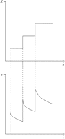
Typical force response to a stepwise and discrete generation of displacement for a visoelastic material. Following each idealized step, the tissue relaxes, and observed force decreases based on the relaxation function.
The relaxation behavior of the tissue can be modeled using a relationship known as the relaxation function [26]. This is an idealized function where a step-input in displacement is imposed on the tissue and is then held constant. The relaxation function describes the force-time behavior of the tissue once the displacement is held. In order to be used as a predictive tool, mechanical properties of the tissue must be understood to provide information to complete the function.
For the remaining activation methods the forces generated during treatment with respect to displacement can be predicted; however, the force with respect to duration of treatment, or time, still remains unknown. Screw-type appliances provide a known displacement, assuming appliance deformation is negligible, to the maxilla, and the resistance of the tissue to this input causes the resulting force generation. Spring-type and magnetic activation methods themselves resist displacement, thus when an appliance utilizing one of these methods is compressed there are already forces present. From Newton's Third Law, which states that the reaction forces between two bodies will be equal and opposite, the forces internal to the tissue during expansion should theoretically be the same as forces produced by the activation method. This statement only holds true if the acceleration of the maxilla is neglected, which in our scenario is a fair assumption since in most cases it will only move approximately 1 cm over the span of several weeks. A great advantage is provided here in that the forces generated during expansion can be predicted. Typical curves for spring-type and magnetic activation methods are illustrated in Figure 3.
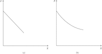
Typical force versus displacement curves for spring-type (a) and magnetic (b) activation methods.
From Figure 3 it can be noted that the curve for the spring-type activation method is shown as linear while the magnetic curve is not.
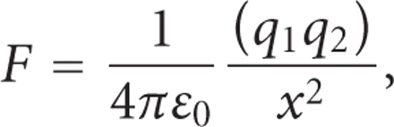
This arises from the fact that the force between two magnets used in repulsion is proportional to the distance separating
them squared, as illustrated in (1). The relationship shown is only for two point charges and as such is a simplification of the physical situation with two
magnets which would involve more extensive analysis; however, it serves as an aid in understanding the force-displacement
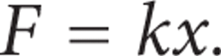
behavior between charged objects [28]. Linear springs will show a linear relationship between force and displacement as shown in (2) and graphically in Figure 3. This may not always be the case as other force-displacement spring relationships may be observed [29].
When considering the force-time behavior during treatment, the general shape of the curves may not be the same as those in Figure 3. The rate at which the force decreases over time is entirely dependent on the resistance of the patient's tissue to deformation. For a patient with very little resistance to expansion, or greater tissue relaxation, the force will decrease faster than if the tissue were to have a higher resistance. As such, accurate prediction of the force-time behavior will be highly patient specific and will require knowledge of the suture properties.
SMA activation methods have not yet been discussed since SMA technology has only been implemented in screw- and spring-type configurations. As such, the force application trends that have been discussed previously for these methods will also apply to SMA appliances, differing only by force magnitude. Since the theory regarding SMAs is highly involved, only a brief discussion will be included here. More detail on this subject can be found in texts such as Physical Metallurgy Principles or in papers such as one by Abramov which discusses functional properties of treated Ti-Ni-Nb alloys [30, 31]. It should be understood that in order to harness the properties of SMAs, the material must be heated into the austenitic region from the martensitic to allow for interface movement or twinning of the materials structure. The human mouth temperature is approximately 37°C which allows clinicians to take advantage of the twinning process that alters material stiffness. Since stiffness is defined as the force required for a unit displacement, it can be seen that the force magnitude applied during expansion will be different when using SMAs over conventional materials, such as stainless steel, when in the patient's mouth.
4.3. General Discussion
Considering the force application of each activation method, a more general discussion of each expansion approach including topic such as clinical implications, ease of activation, and size can be conducted.
4.3.1. Screw-Type Appliances
The greatest advantage of screw-type appliances is arguably the simple and well-understood mechanics [32]. For a given amount of screw rotation there is a corresponding amount of thread-pitch-dependant expansion. This allows clinicians to prescribe a given number of activations to achieve specific expansion between patient visits. Screw-type appliances can easily be designed to be compact and light-weight which is a significant advantage in a narrow maxilla.
While screw-type activation may be the most popular choice for clinicians, it also suffers from the greatest disadvantages of the methods considered. As discussed earlier, the maxilla is subjected to stepwise increments of the appliance which in turn causes rapid increases in forces. This may not only be uncomfortable for the patient, but it has also been suggested that high magnitude forces may result in less physiologic expansion of the suture. Of all the activation methods presented, the screw-type appliances will produce the highest forces. Isaacson found that forces as high as 22.5 lbs, or approximately 100 N, were generated during treatment [24]. A possible way to decrease force magnitude is to decrease expansion rate as this would allow for greater tissue relaxation between activations; however, the rapid increase in force level at activation cannot be avoided.
Another disadvantage of the screw-type activation method is that it requires the patient to activate the appliance; thus, treatment results rely heavily on patient cooperation. Ideally, there should be no patient involvement during treatment to achieve desired results and to remove as much inconvenience to the patient as possible.
4.3.2. Spring-Type Appliances
Continuous force application throughout treatment is an advantage of spring-type activation methods. This minimizes the number, and amplitude, of rapid force increases exerted on the tissue which may lead to more physiologic expansion and increased comfort. Also, force-displacement behavior can be predicted since it is not patient specific. Lastly, patient involvement is eliminated which provides increased patient convenience and improves treatment results.
One disadvantage of the spring-type activation method is that the force output of the device is inversely proportional to expansion. As the deformation of the spring element decreases, the force output will also decrease. This may require intermediate activations to maintain the necessary force magnitude to cause expansion. The ideal situation would see an appliance that could induce expansion with a single low-force activation, meaning that force would need to be independent of displacement and remain constant during treatment.
Another disadvantage of spring-type mechanisms is that they are structurally weak in the directions transverse to expansion. While the device is stable and predictable in the direction of expansion, these mechanisms in general lack the ability to resist forces in the other directions. This disadvantage is more indicative of wire spring-type mechanisms than of coil springs, since the latter typically have an additional structural member providing support. If the spring undergoes unwanted deformation during treatment, then this may have an adverse effect on results. Lastly, spring appliances are inserted into the mouth in a preloaded state. From a design safety perspective this should be avoided since a failure would allow the spring to release and possibly harm the patient.
A device that does not contain an expansion screw or spring is one put forth by McSurdy Jr. in U.S. patent application 20070178421 [33]. Here, the author presents a method of expansion whereby a set of removable appliances, similar to a mouth-guard, are used to incrementally widen the maxilla. Each subsequent appliance is wider than the previous which causes expansion. As a result, this method of expansion does not require patients involvement through continuous activation but does require that they are consistent in wearing the appliance. This device relies on the restoring force characteristic of a spring while showing the discontinuous pattern indicative of screw-type devices. Hence, this procedure will suffer from the same disadvantages as the ones aforementioned for screw- and spring-type activation methods.
4.3.3. Magnetic Appliances
The use of magnets for the purpose of expanding the maxilla is a technique that has been attempted by clinicians such as Darendeliler et al. [10]. When used in the repulsive configuration these types of appliances have many of the same advantages and disadvantages of spring-type appliances. Magnets are able to produce a continuous force without any additional adjustments by the patient. One advantage that magnetic appliances have over spring-type ones, primarily wire spring devices, is that they can be made to be more structurally stable in all directions which will aid in preventing undesirable results.
When using magnets in repulsion, the force output will be inversely proportional to the distance between the magnets. Thus, as the magnets move further apart during treatment the force will decrease in magnitude. As with spring-type appliances this means intermediate adjustments may be required to achieve the desired expansion. One pair of magnets may be used throughout the treatment, but in order to maintain a force level large enough to still produce expansion, the initial forces will need to be significant. Using this alternative may produce less physiologic results due to the high forces exerted on the tissue.
4.3.4. Shape Memory Alloy Appliances
Ni-Ti has been found to be used by clinicians in a coil or wire spring-type method for maxillary expansion [11, 12]. Though the SMA produces more physiologic forces, both in magnitude and in relation to displacement, these devices still suffer from some of the disadvantages of conventional springs. Wire spring devices lack structural stability in the directions transverse to the direction of expansion. Also, a device presented by Darendeliler and Lorenzon, which used a Ni-Ti coil spring, showed that during expansion the force decreased from 800 g (7.85 N) to approximately 400 g (3.92 N) [11]. Though this may not be as significant as the decrease seen with conventional materials, it still shows a 50% decrease in force with displacement.
A device presented by Wichelhaus et al. utilizes Ni-Ti in a combined expansion screw and spring application [13]. The force versus deflection curve shows an improvement from conventional screws as it does not involve the large force jumps seen previously; however, a tensile testing machine was used to gather data which is not a true physical representation of the treatment. As such, while results show promise compared to other screw-type appliances, future testing in a more physically representative environment would be necessary to show the true behavior during treatment. Again, as with all screw-type mechanisms, the patients are significantly involved in this treatment and must activate the screw themselves. Also, the force magnitude reached by this appliance shows forces ranging between 15 N and 20 N which are large compared to other Ni-Ti appliances [11].
Ideal forces to achieve palatal expansion remain unknown. Isaacson pointed out that RME appliances anchored to teeth should produce heavy forces designed to produce minimal tooth movement, while allowing bone repositioning [24]. Lower force magnitudes have been promoted as more physiologic, but when anchored to teeth may cause undesirable tooth movement. Bone anchored appliances may allow lower more physiologic forces for midpalatal suture separation without unwanted tooth movement.
5. Conclusions
From the review conducted in this paper, it was found that the maxillary expansion activation methods in use today could be grouped into four categories: screw-type, spring-type, magnetic, and SMA activation. The following conclusions can be drawn regarding the activation methods in use today.
-
(i) Notwithstanding their popularity in clinical practices, screw-type activation proved to have the most disadvantages in that it requires vast amounts of patient involvement and it induces large magnitude and discontinuous forces
-
(ii) Spring-type appliances showed improvement on screw activation in that they provide continuous force application. Disadvantages of this method are that the force level is dependant on the displacement of the expander, wire springs lack structural stability, and the device is not necessarily fail-safe in case of detachment.
-
(iii) Repulsive magnetic force application suffers from the property that force will decrease as the magnets displace further apart; however, this method can provide low-level forces that are continuous over the displacement.
-
(iv) SMA technology may provide more physiologic force-displacement characteristics than other methods; however, when used in conventional ways such as with a screw or spring, the method still suffers from many of the disadvantages seen with conventional materials.
-
(v) The soft tissue behavior of the sutures when exposed to loading can be used to provide insight into which activation method may provide the most physiologic expansion.
It is clear from the conclusions of this paper that there are more improvements that can be made to maxillary expansion activation methods. Methods that can provide lower levels of displacement-independent forces while requiring no patient involvement would be ideal. Future work should be concerned with determining an ideal force range for maxillary expansion, which would include modeling of the suture, to provide more insight into the design and development of new appliances.
- Received October 29, 2009.
- Accepted May 11, 2010.
- © 2010 D. L. Romanyk et al.


