


Device and materials for in vitro evaluation of forces developed to teeth and periodontal structures during dental practices
- Fotios D Palamidakis1
- Athanasia Panou2
- Kyriaki G Papadokostaki2
- George Leontakianakos3
- Vassilis N Stathopoulos3
- Evangelos G Kontakiotis1
- 1Department of Endodontics, Dental School, University of Athens, Athens, Greece
- 2Department of Physical Chemistry, IAMPPNM, National Center for Scientific Research “Demokritos”, Agia Paraskevi Attikis, Greece
- 3General Department of Applied Sciences, School of Technological Applications, Technological Educational Institute of Sterea Ellada, Psahna, Greece
- Evangelos G Kontakiotis, Department of Endodontics, Dental School, University of Athens, 2 Thivon Str, 115 27 Goudi, Athens, Greece. Email: ekontak{at}dent.uoa.gr
Abstract
This study aimed at providing a gauge device (Ekontak et al Gauge K-Device) in order to analyze the forces applied to teeth and periodontal tissues during dental practices in vitro. This force gauge device can be used in the investigation of the possible defect generation to tooth structures when overloaded forces are applied during dental procedures in vitro. Ekontak et al Gauge K-Device consists of three units: the specimen’s holder, a high-performance digital force gauge, and the support frame. The holder was fabricated by an Al alloy providing a steady detachable attachment between the specimens and the force gauge’s pin connector. The clinical simulation was achieved with the use of a proper silicone material, selected to provide similar elastic behavior with the human periodontal ligament and to join the teeth inside a solid matrix of an acrylic resin. The digital force gauge is a high-speed collection and recording (1000 Hz) product coupled with data recording software. The forces developed to 15 specimens’ root canals during lateral condensation and vertical compaction of cold gutta-percha obturation procedures were monitored, saved as graphs, CSV, and excel files and presented over time. The forces developed during vertical compaction (mean maximum force per obturation circle = 13.22 N) were more excessive than those during lateral condensation (mean maximum force per obturation circle = 10.14 N). In conclusion, Ekontak et al Gauge K-Device is provided as a modern gauge device, capable of performing clinical simulation in vitro, under the terms of its protocol.
Introduction
During endodontic treatment, forces are applied inside the root canal. Lateral condensation and vertical compaction of gutta-percha (GP) are examples of practices involving pressure and forces’ application inside the root canal system.1⇓–3 The intensity, the direction, as well as the duration of these forces vary.1⇓–3 These forces may have an impact on dental structures, such as dentine, enamel, and cementum, developing defects, which might later propagate into fractures with severe clinical consequences.1⇓⇓⇓⇓–6
A “cushion effect” over these forces exists by the complex human periodontal ligament (PDL)–adjacent alveolar bone (AAB). PDL tends to act as a shock absorber during mastication, although the tooth’s movement reaction to external force application follows a rather complicated pattern.7⇓–9
Especially during obturation techniques, according to Schilder,10 pressure is needed in order to adapt GP to the walls and to achieve a tridimensional and hermetic filling of the root canal. The fact that GP performs more or less like a noncompressible material results in an increase of the hydraulic forces applied inside the root canal.11,12
Whenever excessive condensation or compaction forces are applied inside a root canal, root fracture, dentinal defects, cracks, or even complete root and tooth splits may occur.4 Similar harmful impact on the PDL and the AAB may exist too, but less often and under extended gauges.1⇓⇓–4,13
A number of investigators have studied this phenomenon using different types of gauge devices.1⇓⇓–4,13⇓⇓⇓⇓⇓–19 Among them, Blum et al.1⇓–3 evaluated the forces developed during warm vertical compaction technique and analyzed the wedging effect. Saw and Messer17 demonstrated that root canal obturation is the major cause of vertical root fracture because of the wedging effect generated in the canal by the compaction strain. Ricks-Williamson et al.18 studied the stress induced by obturation with finite-element analysis. This study’s experimental measurements showed that vertical rather than lateral forces developed are more detrimental throughout the root canal. Other authors experimented using different testing methodologies, such as the Instron testing machine20,21 and photoelastics.22 Shemesh et al.,5 in 2010, correlated the forces applied during obturation procedures with the occurrence of dentinal defects.
Up to date according to our knowledge, no studies have been reported to deal with the materials’ choice in order to simulate in vitro the tooth and periodontal system.
The aim of this study was to provide a gauge device, Ekontak et al Gauge K-Device (EGK), in order to measure, continuously record with fast data acquisition, and analyze the forces developed inside the root canal during the different steps of endodontic treatment and especially during obturation (Figure 1). EGK applies to the name of the simulating device created and indicates the research team that inspired and constructed it.
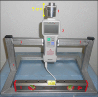
The EGK: 1 refers to the holder, 2 to the gauge device, 3 to the stands’ elbow brackets, 4 to the support frame, and 5 to a level indicator.
EGK: Ekontak et al Gauge K-Device.
Additional aim was the investigation and use of a proper material for the preparation of the tooth specimens. Therefore, in order to simulate in vitro the tooth and periodontal system, a dental elastic material with similar mechanical properties (modulus of elasticity) with the human PDL has been investigated.
Materials and methods
Device
The created gauge device (EGK) consisted of three units: the holder containing the specimens, a high-performance digital force gauge, and the support frame (stand). It is supported by a personal computer (PC) for data acquisition and storage through a USB 2.0 connection and appropriate software that is described later.
The holder was fabricated by an Al alloy (Al 7075) and is screwed on top of the gauge pin connector (Figures 1 and 2). The purpose of the holder is to provide a steady detachable attachment between the specimen and the force gauge’s pin connector.
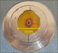
The holder (No. 1), supporting a specimen. No. 2 refers to the acrylic resin and the arrow (No. 3) shows the silicone material. The tooth’s root is attached inside a thin phase of the silicone material.
Moreover, the holder was used as a mold in order to embed the 15 tooth specimens used for the purpose of this study.
The digital force gauge (Digital Force Gauges by Imada: ZP series, Imada Co., Ltd, Japan) is a high-speed collection and recording (1000 Hz) product (Figure 1). The maximum permissible compression or tensile force is 50 N, while the resolution is 0.01 N.
The measuring unit is coupled with data recording and transmitting software, ZP recorder. It is a force data analysis software designed to control and interface with ZP Series (USB) force gauges. ZP recorder is suitable for high-speed sampling, processing 1000 data per second, not just the peak of them, and generates a graph with statistics plus an excel complete format.
The device and holder are mounted and supported by a steel framework. It is compact, bench top designed, able to securely accommodate the holder and the force gauge (Figure 1).
Specimens
The specimens were prepared using the holder as a mold, in a way that they actually resemble the in vivo structure, tooth—human PDL—AAB.
The teeth selection
For this study, 15 freshly extracted, human, single-rooted mandibular premolars of similar size, shape, and root canal anatomy were selected. The extractions of the teeth were atraumatic and most of them for periodontal reasons. All roots were examined with an optical microscope under a 3× magnification (Protégé Plus, Global Surgical, Inc., St Louis, MO, USA) to exclude cracks. The teeth were stored afterward at 37°C in purified water, for a maximum period of 3 weeks, until obturations completed. The patients’ age varied between 45 and 55 years.
Prior to any specimen preparation or testing, all the teeth were subjected to a two-dimensional, mesiodistal and buccolingual, digital radiographic examination (iOX2 CCD sensor, pixel size 25 × 25 µm; Fimet OY, Monninkylä, Finland) using an IntraOs 70 dental X-ray unit (Blue X, Chicago, IL, USA) under standardized conditions. The exposure parameters were 70 kVp, 7 mA, with an exposure time of 0.06 s. Only those teeth that contained one straight and round root canal with an average radiographic diameter of 0.7 mm between the buccolingual canal walls at a distance of 10 mm away from the apex were selected for this study. During this procedure three teeth were disqualified and replaced by suitable ones.
Afterward, all teeth’s crowns were sectioned using a low-speed saw (Isomet 11-1180, Buehler Ltd, Evanston, IL, USA) under running tap water, perpendicular to the long axis of the root at the level of 12 mm away from the apex and rinsed thoroughly with sterile saline.
PDL’s simulation material
Concerning the simulation of the PDL, a proper material among five different impression materials was studied in terms of their mechanical properties. The five materials, tested for the determination of their Young’s modulus, were the following: Gumak Condensation Light (Acteon Corporation, Norwich, UK), Gumak Condensation Putty (Acteon Corporation), Aquasil Ultra Monophase (Dentsply-Maillefer, Ballaigues, Switzerland), Splash Light (Discus Dental, LLC, Copenhagen, Denmark), and Splash Putty (Discus Dental, LLC).
Material samples with dimensions of 1 × 20 mm2 and thicknesses of 450–570 µm were tested with a tensile tester (Tensilon UTM-II-20, Toyo Balwin, Co. Ltd, Japan). Stress–strain tests with an initial gauge separation of 10 mm were performed at a constant rate of elongation of 10 mm/min, at T = 23 ± 2°C and relative humidity 63 ± 4%. To check the repeatability, at least five samples from each material were measured.
A thin film of the selected dental silicone material of thickness approximately 250 µm, close to the human PDL thickness,23 will resemble the PDL’s absorber role joining the teeth inside a resin block (Caldofix-2 Resin, Struers A/S, Ballerup, Denmark), supporting the silicone material with the tooth’s root solidly (Figure 2).
Specimens’ preparation
The holder was used as a mold where Caldofix-2 Resin was cast. The tooth root was covered by a plastic paraffin film (Parafilm, Pechiney Plastic Packaging Company, Richmond, VA, USA). The stretched paraffin film of thickness about 250 µm had good adhesion on the tooth root substrate and provided excellent sealing against liquid, that is, resin, intrusion. The film-covered tooth root was placed inside the epoxy resin up to 2.0 mm coronal height and left until resin’s setting. The paraffin film was covered with petroleum jelly (100% pure Vaseline Petroleum Jelly, Unilever Ltd, Epping, NSW, Australia) in order to avoid adhesion of Caldofix-2 Resin. Then the tooth root was removed leaving in the resin its imprint and was uncovered from the protective film. The imprinted cavity, after being thoroughly cleaned, was covered by the selected dental silicone material to resemble the PDL. The tooth’s root was protected in the same method but top–down to 2 mm coronally and fitted back in the cavity.
As soon as the silicone reaches the setting time, the excessive quantity of the silicone material as well as the protective film were removed and the specimen (resin + silicone + tooth’s root) was separated from the holder for another specimen to be prepared.
Root canal obturation
The root canals’ chemomechanical treatment and obturations were performed by a specialist in endodontics.
The roots were prepared up to F3 size NiTi Protaper finishing file (Dentsply-Maillefer), according to the manufacturer’s protocol,24 before placing in the device through the holder.
In all cases as soon as a specimen got attached inside, the holder of EGK and the obturation were ready to start; the gauge device was switched on to the record mode and the data recording was initiated by a PC-controlled ZP software. The data were transferred through a USB 2.0 connection to a PC. The data recordings were saved as CSV files, excel files, and graphs.
In all cases, the root canal was filled with the lateral condensation technique according to the protocol,5 using a size 40 GP master cone (Roeko, Coltène/Whaledent, Inc., Cuyahoga Falls, OH, USA) and AH-26 (Dentsply DeTrey, Kostanz, Germany) was used as the root canal sealer. A size C spreader (D1 diameter 0.3 mm, 0.04 taper) (Dentsply-Maillefer) and size 25 standardized GP cones (Roeko, Coltène/Whaledent, Inc.) were used for the lateral condensation according to the protocol. The coronal GP was removed with a heated plugger of 0.5 mm diameter (Dentsply-Maillefer) with the less possible force applied. Finally, coronal vertical compaction was performed using a cold, manual plugger 3/4 (Dentsply-Maillefer).
All 15 specimens were subjected to the same experimental procedure.
Results
The silicone material selected for the PDL’s simulation
The results from the five materials, tested for the determination of their Young’s modulus, are shown in Figure 3 in the form of stress–strain curves. In the same figure, the Young’s modulus E determination is shown and the determined values for each material as well.
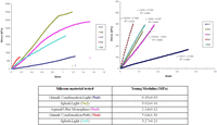
Stress–strain curves of the PDL-resembling materials tested.
PDL: periodontal ligament.
Among Gumak Condensation Light, Gumak Condensation Putty, Aquasil Ultra Monophase, Splash Light, and Splash Putty, the first was the material selected.
According to the values reported in the literature, Gumak Condensation Light’s physical properties concerning the Young’s modulus E, measured as 0.45 ± 0.03 MPa, were by far closer to that of the human PDL (0.12–0.96 MPa depending on the load forces applied during tests) reported by Yoshida et al.7 and within the range of valid values reported in other works.25⇓–27 According to these results, Gumak Condensation Light was proven the most appropriate material and was used in the specimens’ preparation.
The evaluation of the forces applied during the obturation procedures
Two representative graphs from the recording of the forces applied in each obturation procedure are shown in Figure 4(a) and (b). In all 15 cases, forces ranged up to a maximum of 24.07 N of compression force. The forces developed during vertical compaction (mean maximum force per obturation circle = 13.22 N, standard deviation (SD) = 6.85 N) were more excessive than those during lateral condensation (mean maximum force per obturation circle = 10.14 N, SD = 4.13 N). Tensile forces were also recorded.
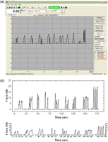
Two graphs recorded during the obturation of two root canals with the use of EGK: (a) The computer’s screen icon during the device’s operation during a root canal obturation. (b) A diagram generated after a root canal obturation (the lateral condensation’s seven circles and the vertical compaction’s last phase are highlighted).
EGK: Ekontak et al Gauge K-Device.
Discussion and conclusion
It has been confirmed that root canal preparation with rotary nickel–titanium instruments can damage the dentine and create defects on root canal walls.4 Additionally, filling the prepared root canal with lateral compaction could induce damage to the roots.28 The forces that the endodontic instruments (pluggers, heaters) apply to teeth’s root canals during endodontic treatment may have an impact on the dentinal structure and provoke certain abnormalities, such as cracks, craze lines, or even root fractures and splits.4,5
The forces’ strength, time of application, and analysis have been tested and measured by some authors in the past.1⇓–3,5,13⇓⇓⇓⇓⇓⇓⇓⇓–22 Blum et al.,1 in 1997, introduced a gauge device (Endographe) used in five experimental in vitro studies.1⇓–3,5,13 These studies analyzed the forces developed during different obturation procedures and correlated the measurements with an impact on the dentinal structures.
In this study, the EGK device proved able to achieve a fast acquisition of force values as developed during obturation. Our primary results’ analysis concerning the forces’ application range during obturation procedures performed by a specialist in endodontics revealed that the forces generated were within the limits of those obtained by the literature in similar researches.1,5 Further investigation is needed in order to correlate the forces’ application exact range with the possible dentinal defects generation.
Whereas one of the issues of the study of Shemesh et al.5 in 2010, which used a simulation of the Endographe, was the absence in vitro of a material acting as the human PDL, in this study, five different polymers were tested for this purpose and the more suitable among them (Gumak Condensation Light) is proposed, based on the results of the tensile testing.
This study presents EGK with a procedure protocol as a clinical simulation device for future attempts to measure and analyze the forces applied during endodontics and other aspects of modern dentistry practicing. While EGK’s function is limited on measuring only the axial components of the forces’ application that are found to be the most detrimental,18 it is an improved device regarding the speed of the information transmitted through the digital gauge device to the computer, which reaches the 1000 data per second and the high resolution, that is, 0.01 N. EGK is 10-fold faster, providing a full curve of data during a single plugging stroke that usually lasts a few milliseconds. The high data acquisition capability is rather useful especially during the last step of obturation when periodical vertical compaction forces are applied in sets of ~20 or more. At the same time, the tensile forces can also be measured by the EGK. However, this was not an object of this study.
Furthermore, the protocol of this clinical simulation device includes a certain specimen preparation, resembling in vitro the structure tooth—human PDL. This approach has not been dealt in the literature according to our knowledge. Thus, PDL’s role is given by an extra thin phase (approximately 250 µm) of a dental silicone impression material (Gumak Condensation Light), supported solidly by a certain epoxy resin (Caldofix-2 Resin).
The real-time in vivo situation involves parameters, such as the adjacent teeth presence or absence, the type of the tooth’s reconstruction as well as the extent of the access cavity preparation and the tooth’s exact periodontal condition, that complicate the evaluation of the phenomenon tested. However, under the terms of this device’s protocol, many of these parameters were isolated successfully, focusing on the parameter forces’ application to teeth and periodontal structures during obturation in vitro.
The educational perspectives of the device and the spread of the results of the scientific project using the EGK fall within the future aims of the authors. Therefore, it is under experimental development and test the investigation of the possible defects to tooth structures when overloaded forces are applied during dental procedures in vitro, using EGK device and the proposed protocol. The high data sampling rate per millisecond leaves no blind time range during plugging.
However, the EGK device and protocol is an extra tool toward off-line analysis of a given obturation procedure as well as the pretreatment steps. The use of X-rays is indispensable for an obturation procedure performed in a state-of-the-art way.
In conclusion, EGK is provided as a modern gauge device, capable of performing clinical simulation in vitro, under the terms of its protocol.
Article Notes
-
Declaration of conflicting interests The authors declare that there is no conflict of interest.
-
Funding This research received no specific grant from any funding agency in the public, commercial, or not-for-profit sectors.
- © The Author(s) 2013


