- Institution: Stanford Univ Med Ctr Lane Med Lib/Periodical Dept/Rm L109
- Sign In as Member / Individual
Splice Variants of GPCRs
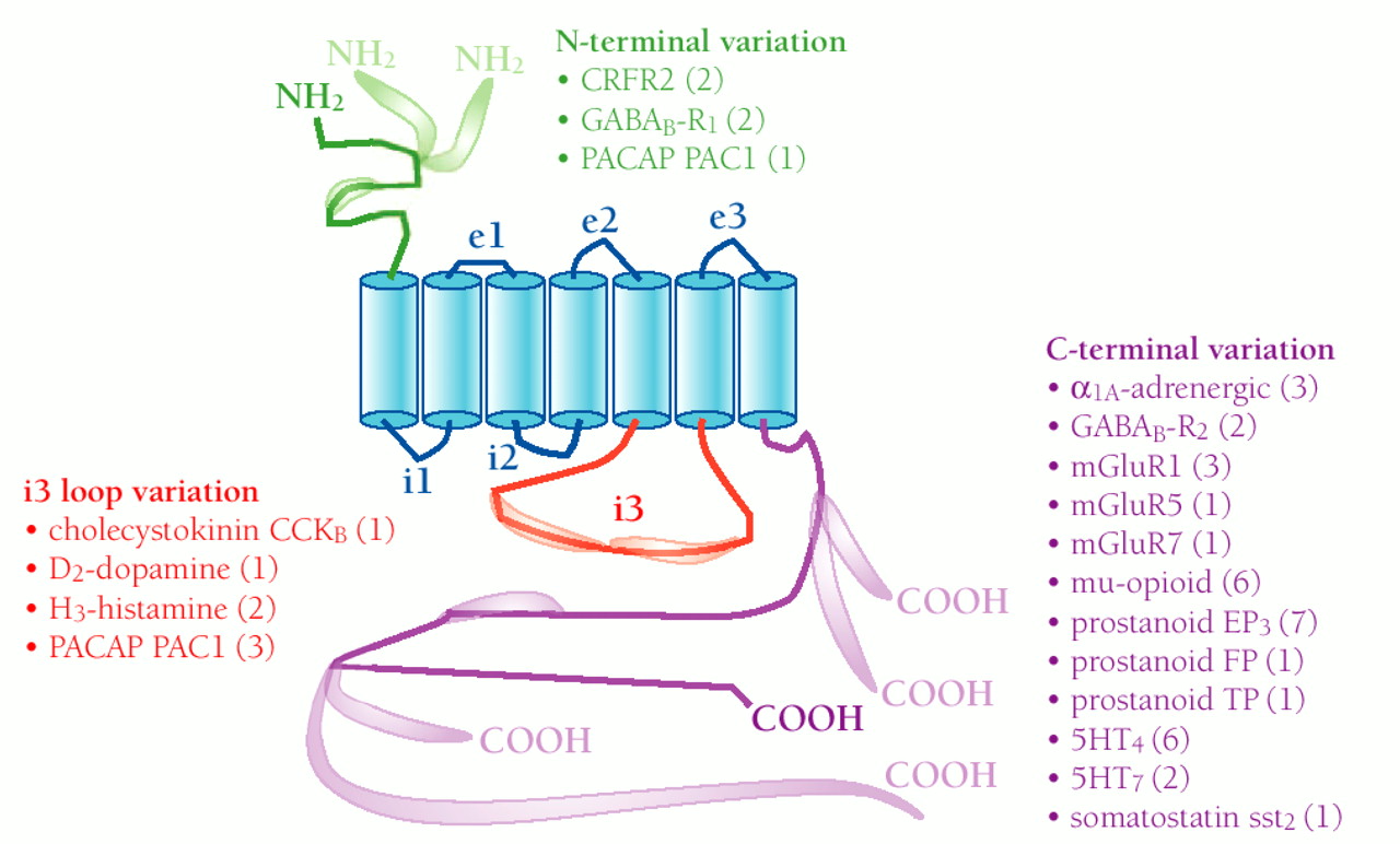
Schematic simplification of the GPCR structure and summary of three classes of human GPCR splice variants. The seven transmembrane domains are shown as blue cylinders; the remaining canonical polypeptide sequence is shown as colored string: extracellular loops (e1, e2, and e3), and two intracellular loops (i1 and i2) are shown in blue; the N-terminal sequence is green; intracellular loop i3 is red; and the C-terminal sequence is purple. Alternative splice variants, including those that represent alternative N and C termini, are indicated by the pod-like expansions superimposed upon the string. Numbers in parentheses next to receptor names indicate how many splice variants exist in addition to the first receptor cDNA sequence cloned. Human sequences verified by two independent labs or approaches are included. References for each of the splice variants are available in the online version of this article.
This Article
-
MI June 2001 vol. 1 no. 2 108-116

