- Institution: Stanford Univ Med Ctr Lane Med Lib/Periodical Dept/Rm L109
- Sign In as Member / Individual
Signaling with Phosphoinositides: Better than Binary
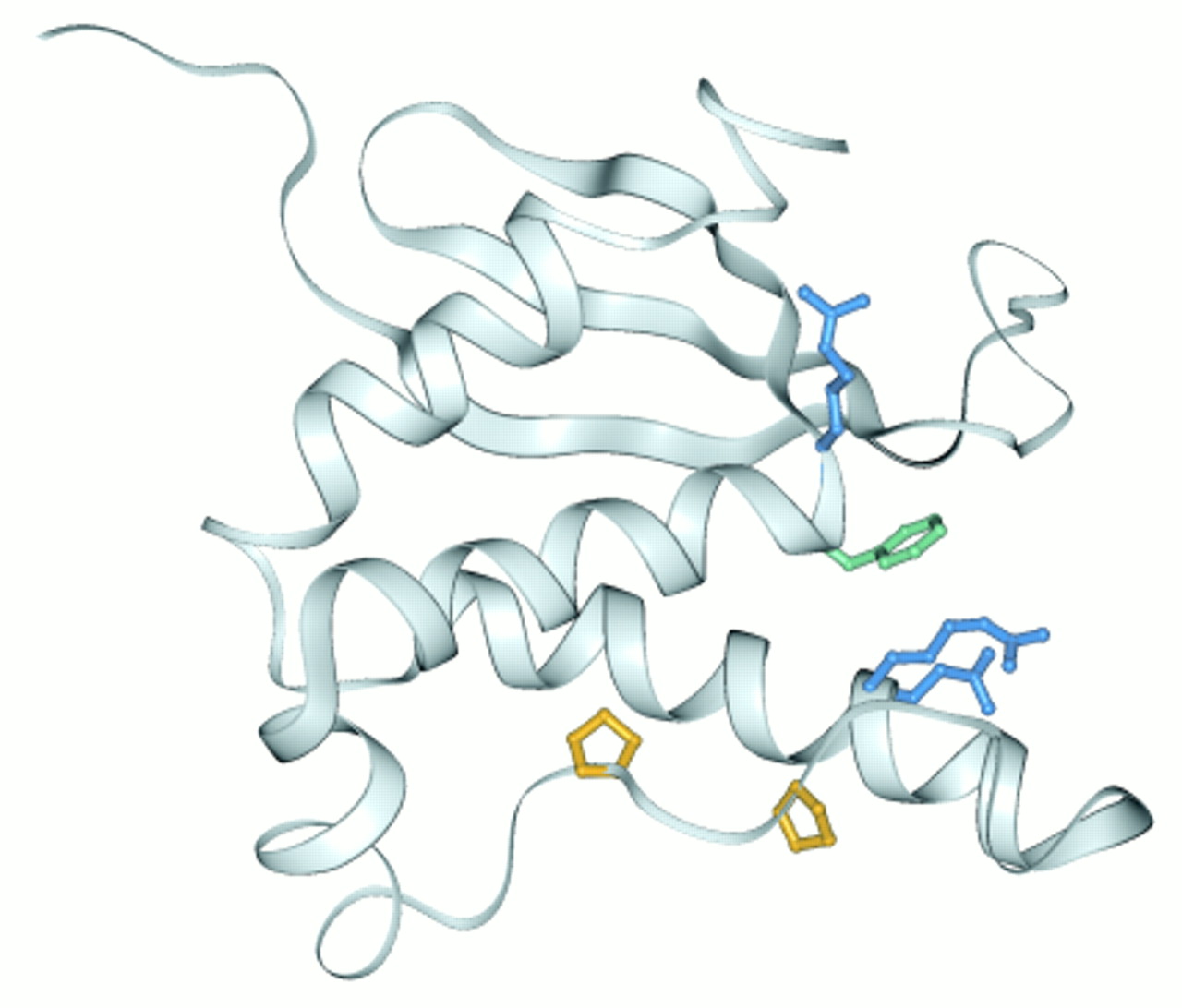
Figure 5.
Structure of the PX domain of p47phox. The backbone ribbon depicts the three-stranded antiparallel β-sheet followed by a bundle of three α-helices and short 3/10 helix (39). A pair of proline side chains is shown in yellow to indicate the polyproline type II helix recognized by the SH3 domain. The predicted PI binding elements (3) that form an accessible basic pocket include the arginine (blue) and phenylalanine (green) side chains of the RRφ motif that precedes the first α-helix and pR+ motif in the second α-helix. (See text for details.)

