- Institution: Stanford Univ Med Ctr Lane Med Lib/Periodical Dept/Rm L109
- Sign In as Member / Individual
Neuropathic Pain: The Paradox of Dynorphin

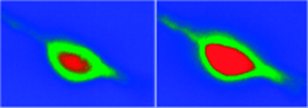
Figure 2.
Dynorphin A(2–17) mediates an increase in intracellular calcium ion concentration in rat cortical neurons. The pseudocolor image shows the basal fluorescence of a typical neuron loaded with the calcium-sensitive fluorophore fluo-3 (left) and the same cell after the administration of 30 μM dynorphin A(2–17) to the bathing medium (right). Dynorphin A(2–17) induces a significant increase in the fluorescence of fluo-3 (expanded red area); the maximum fluorescence is reached after 1 min.


