- Institution: Stanford Univ Med Ctr Lane Med Lib/Periodical Dept/Rm L109
- Sign In as Member / Individual
Networking with AKAPs

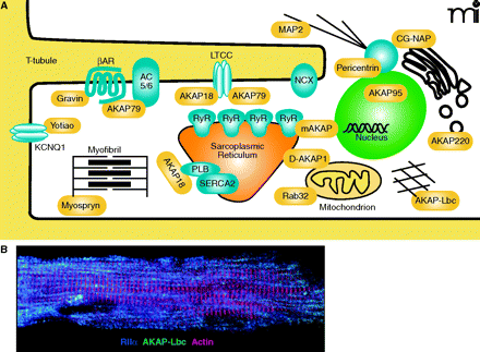
The coordination of PKA signaling events in a cardiac myocyte by localized scaffolding of AKAPs. A. In a cardiomyocyte, multiple AKAPs (yellow) coordinate physiologic and pathophysiologic signaling events, including excitation-contraction coupling, hypertrophic remodeling, gene transcription, and oxygen homeostasis. B. Adult mouse cardio-myocytes were fixed and incubated with antibodies against RIIα (blue) and AKAP-Lbc (green), and actin was visualized by phalloidin staining (red). Cells were then imaged using immunofluorescence microscopy. RyR, ryanodine receptor; βAR, β-adrenergic receptor; NCX, sodium-calcium exchangr ; CG-NAP, centrosome- and golgi-localized PKN-associated protein; MAP2, mitochondrial-associated protein 2 ; LTCC, L-type calcium channel; AC5/6, adenylyl cyclase 5 or 6; PLB, phospholamban; SERCA2, Sarcoplasmic-endoplasmic calcium pump 2.


