Age-related Macular Degeneration: Genetic and Environmental Factors of Disease

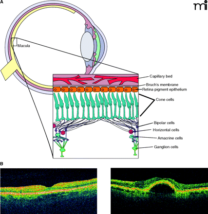
Figure 1
The retina revealed. A. Diagrammatic representation of the retina. Inset shows the several types of neuronal cells within the macula, the retinal pigment epithelium (RPE), and choroid. B. Two optical coherence tomography images of a normal (left) and AMD retina with pigmented epithelium detachment (PED) (right).


