Age-related Macular Degeneration: Genetic and Environmental Factors of Disease

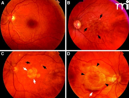
Figure 2
Fundus images from patients representing several AMD subtypes. A. Normal macula. B. Macula with confluent soft drusen (black arrows), a hallmark of early AMD. C. Macula of dry AMD with soft drusen (black arrows) and geographic atrophy GA (white arrows). D. Macula of choroidal neovascularization (CNV) or wet AMD with subretinal hemorrhage (black arrows).


