- Institution: Stanford Univ Med Ctr Lane Med Lib/Periodical Dept/Rm L109
- Sign In as Member / Individual
Isoforms of Mammalian Adenylyl Cyclase: Multiplicities of Signaling
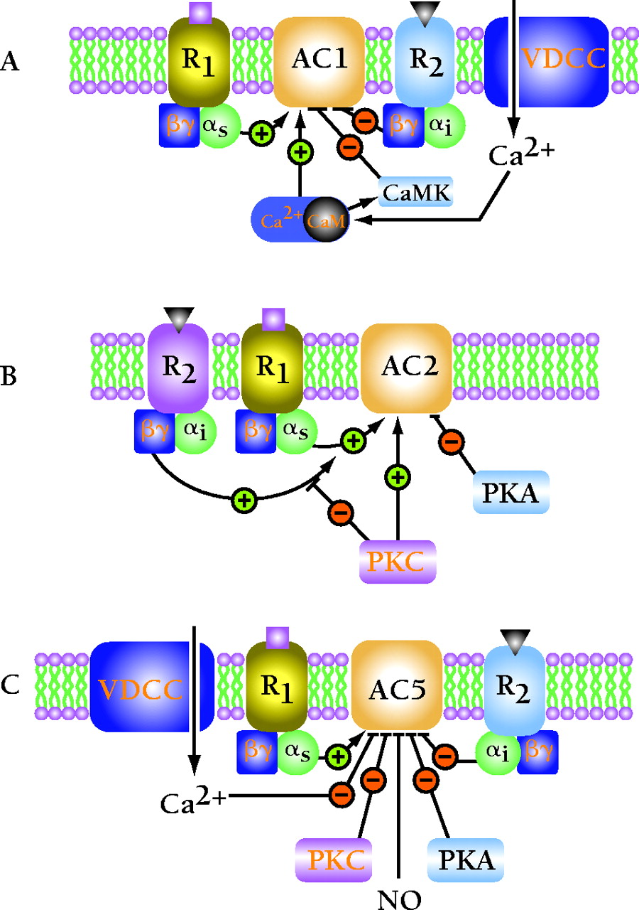
Multiple modes of regulation of adenylyl cyclase isoforms. (A) The pattern of regulation of AC1 as illustrated is representative also for AC3 and AC8. R1 represents a G protein–coupled receptor, such as the glucagon or β2-adrenergic receptor, that couples to the stimulatory G protein Gαs. R2 represents a G protein–coupled receptor, such as the muscarinic M2 or α1-adrenergic receptor, that couples to the inhibitory G protein Gαi. (B) The pattern of regulation of AC2 as illustrated is representative of the regulation of AC4 and AC7. Note that Gβγ regulation of AC2 is dependent on Gαs co-activation and does not activate AC by itself. PKC can use AC as a substrate, resulting in elevation of basal activity and inhibition of the Gβγ superactivation. (C) The pattern of regulation of AC5 is representative also of AC6. (PKA, protein kinase A; PKC, protein kinase C; CaM, calmodulin; CaMK, calmodulin-dependent kinase; NO, nitric oxide; VDCC, voltage-dependent Ca2+ channel.)

