- Institution: Stanford Univ Med Ctr Lane Med Lib/Periodical Dept/Rm L109
- Sign In as Member / Individual
GAF Domains: Two–Billion–Year–Old Molecular Switches that Bind Cyclic Nucleotides

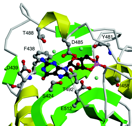
Figure 3.
Detailed view of the cGMP-binding GAF B motif from mouse PDE2A. Shown are six side chains (D485, T488, T492, D439, S424, and E512) that make relatively close hydrogen bonds to the cGMP ligand, either directly or through one of three bound water molecules. In addition, the phenyl side chain of F438 stacks with the guanine ring, and two backbone amides (I458 and Y481) interact with the phosphate group. Beta sheets are colored green and alpha helices yellow. Cyclic GMP is shown in black. Side chains that interact with cGMP are shown in gray. Hydroxyl groups are in red and nitrogen atoms in blue. Water molecules are indicated in light blue.


