- Institution: Stanford Univ Med Ctr Lane Med Lib/Periodical Dept/Rm L109
- Sign In as Member / Individual
A “Wringing” Endorsement for Myosin Phosphorylation in the Heart
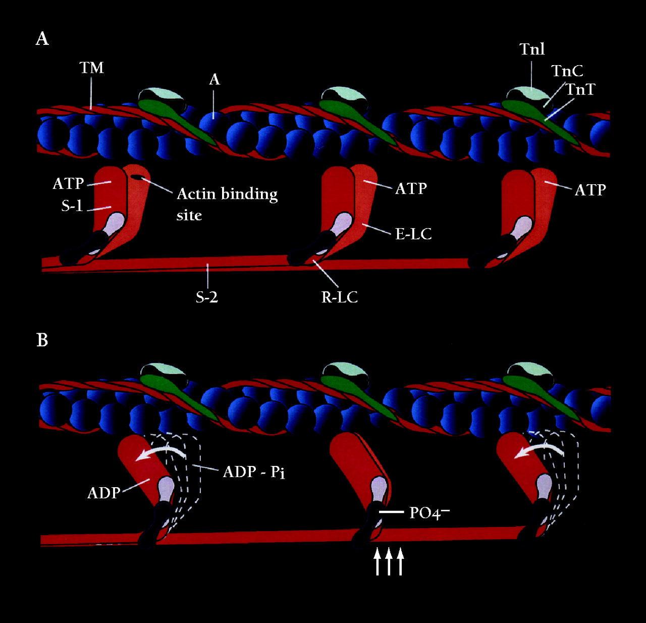
Schematic representation of the contractile and regulatory proteins of the thick and thin filaments of the cardiac sarcomare. A. The actin monomers (A) of the thin filament (blue spheres) and the cable-like tropomysin (TM) molecule (red), as well as the regulatory protein complex consisting of troponin I(TNA)(light blue), troponin C(TNC)(dark green) are shown. The S-I or motor head domain of myosin (red and orange), including the essential light chain (E-LC) (light purple) and the regulatory light chain (R-LC) (purple), is also depicted. S-2, part of the tail domain, oors S-1 to the thick filament.B. The movement of S-1 towards the thin filament proposed to occur following phosphate incorporation(PO4−) by the R-LC is shown(vertical arrows). The force-generating power stroke is energized by the hydrolysis of ATP to its products ADP = P,iand the subsequent release of Pi(curved arrows). Phosphate incorporation by the R-LC may facilitate this force generating event during submaximal Ca2+activation.

