- Institution: Stanford Univ Med Ctr Lane Med Lib/Periodical Dept/Rm L109
- Sign In as Member / Individual
LQT4 Gene: The “Missing” Ankyrin

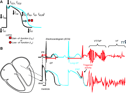
Cardiac action potential, electrocardiogram (ECG), and long QT syndrome (LQTS). A. In LQTS, mutations cause either a gain- or a loss-of-function in INa or IK currents, respectively, that results in prolongation of the cardiac action potential. B. Representative ECG traces in relation to normal atrial and ventricular action potentials. PR and QT intervals, respectively, correspond to the atrial and ventricular activation and repolarization times. Prolongation of the cardiac action potential is reflected as lengthening of the QT interval (blue), an increased vulnerability for the development of early afterdepolarization (EAD), “triggered activity,” and ultimately cardiac arrhythmias [torsade de pointes (TdP) or ventricular fibrillation (VF)]. IKr, rapidly activating delayed-rectifier potassium channel current; IKUR, ultrarapid delayed rectifier potassium current; ITO, transient outward potassium channel current; IK1, inward rectifier potassium channel current.


