- Institution: Stanford Univ Med Ctr Lane Med Lib/Periodical Dept/Rm L109
- Sign In as Member / Individual
The Biology of Caveolae: Lessons from Caveolin Knockout Mice and Implications for Human Disease
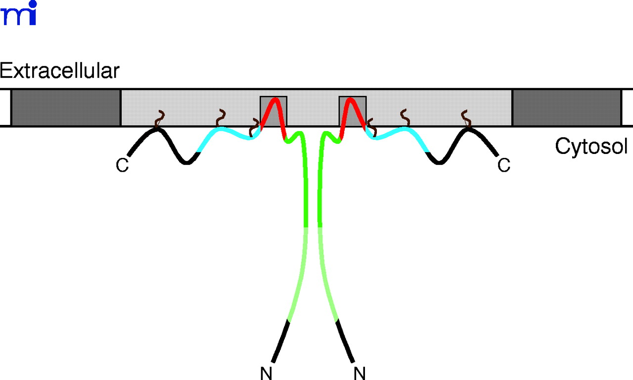
Figure 2.
Topology of caveolin at the inner cell membrane. A dimer of Cav-1 is represented; interprotomeric association is maintained through the so-called oligomerization domain (dark and light green). Association of Cav-1 with the cell membrane is maintained by N-terminal and C-terminal membrane attachment domains (dark green and blue, respectively), the transmembrane domain (red), and three palmitoyl groups (brown). The plasma membrane is indicated in dark gray; cholesterol- and sphingomyelin-enriched membrane is indicated in light gray.

