- Institution: Stanford Univ Med Ctr Lane Med Lib/Periodical Dept/Rm L109
- Sign In as Member / Individual
The gβγ Dimer as a Novel Source of Selectivity in G-Protein Signaling: GGL-ing AT CONVENTION
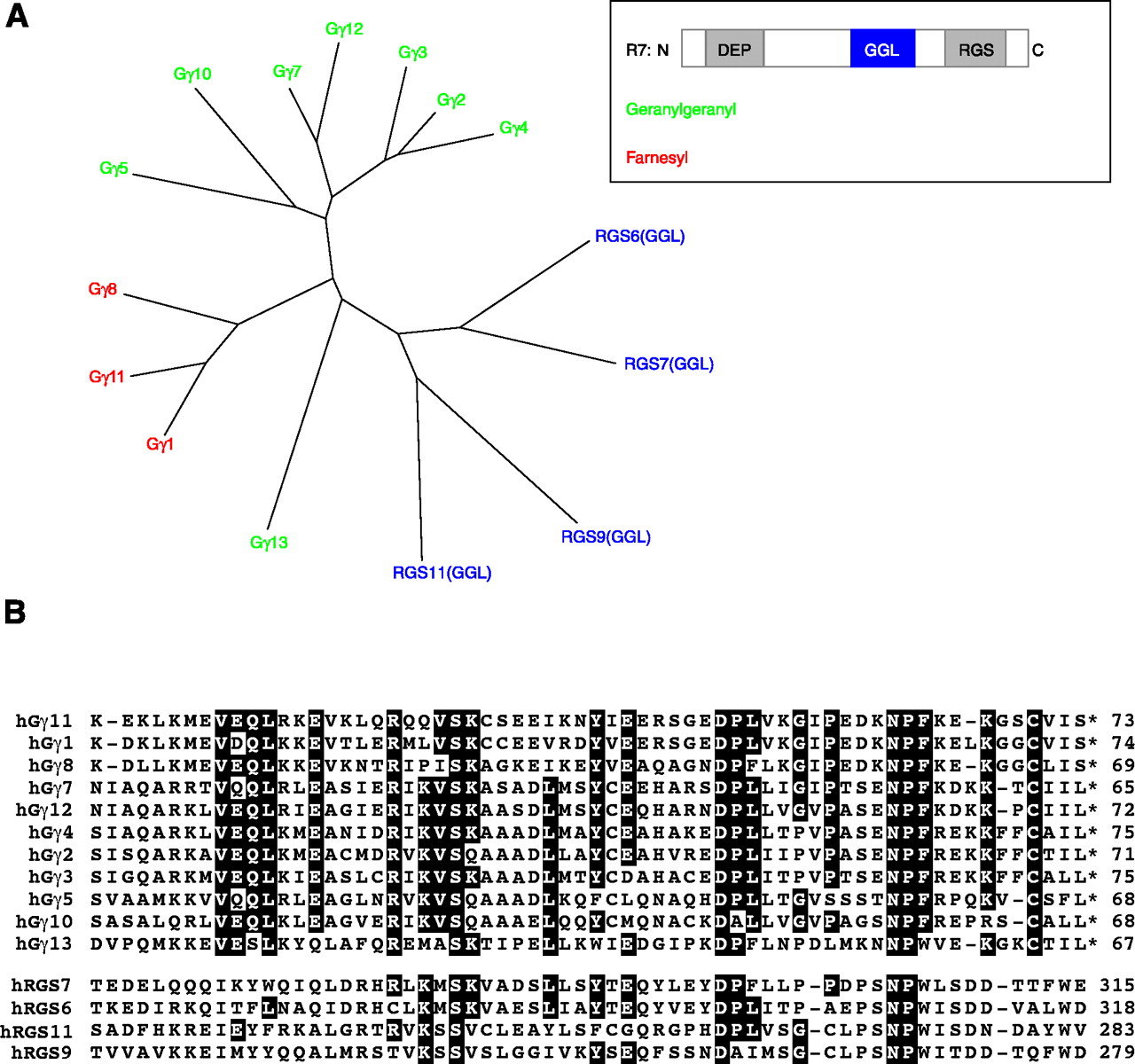
The relationship between the human Gγ subunit proteins and R7 subfamily of RGS proteins. A. An unrooted dendrogram depicts the degrees of similarity among proteins. As shown in the inset, R7 subfamily members contain a DEP domain, GGL domain, and RGS domain; only the GGL domain (blue) sequences were used for the dendrogram analysis. Gγ subunits are post-translationally modified with a geranylgeranyl (green) lipid or a farnesyl (red) lipid moiety. The unrooted dendrogram was generated using TreeView X from a multiple sequence alignment created with ClustalW using default settings. B. Multiple sequence alignment of human Gγ subunits and GGL domains of human R7 subfamily of RGS proteins. Black boxes depict identical amino acids shared by at least 60% of sequences within alignment. Sequence alignment between human Gγ subunits and GGL domains was computed by the Wisconsin GCG Pileup program using default parameters. GenBankTM gi identifier numbers for human sequences used: RGS7, 17380284; RGS6, 31742476; RGS9, 8475983; RGS11, 34452688; γ11, 4758448; γ1, 11386179; γ8, 3023844; γ7, 4826746; γ12, 40254926; γ4, 4758450; γ2, 11277005; γ3, 6912394; γ5, 4885287; γ10, 4758446; γ13, 7706567.

