A CENTURY of ULCER MEDICINES
The drug treatment of peptic ulcer in the 20th century has a most intriguing history. During the first three-quarters of that century, numerous medications were used whose development was based on physiology studies performed in the 1820s, and these ulcer therapies typically acted locally in the stomach. The final quarter of the 20th century saw a true paradigm shift: elucidation of cell receptors and biochemistry of the stomach guided the development of two classes of highly effective, systemic-acting agents. At the same time, an unexpected microbial etiology was proposed, shaking up the prevailing sense of certainty about the origin of peptic ulcers, and introducing still another class of therapeutic agents.
This article looks at the variety of pharmacologic agents used to treat peptic ulcers and related ailments, and the rationales behind their use. It also discusses the technologies that supported their development, and the seminal contributions made by three research scientists whose dedication and initiative transcended all limits. Although focused on 20th century drugs, the story really begins early in the 19th century….
1825: Beaumont Looks into St. Martin’s Stomach
The acidity of stomach secretions and their ability to digest meat were detected in 1783 by the brilliant Italian experimental biologist Lazzaro Spallanzani (1), but it remained for an obscure American army doctor, William Beaumont, to elucidate the functioning of the stomach based on direct visual observation. This major American contribution to the science of physiology came not from Philadelphia or Boston, but from a frontier military outpost in Fort Mackinac, Michigan.
William Beaumont (1785–1853) was a farm boy from Lebanon, Connecticut. Seeking independence, he left his family’s home at the age of twenty-two, with no specific destination in mind. After running a village school in Champlain, New York for three years, he went to St. Albans, Vermont, where he apprenticed to a physician, Dr. Benjamin Chandler, for two years. In 1812, he received his license to practice medicine, and promptly joined the army as an assistant surgeon. He distinguished himself in the War of 1812, both on land and water (Lake Ontario). As a sidelight, his journal provides a harrowing description of the effects of an explosion of 300 pounds of gunpowder, deliberately set off by the British, which killed sixty American soldiers outright, and wounded more than 300. His description of men “mashed and mangled” reminds one of the suicide-homicide bombings of the present (2).
Beaumont left the service in 1815, angry that less experienced men had been promoted over him. He settled in Plattsburgh, New York and successfully practiced medicine there for four years. His former army friends persuaded him to rejoin, and in 1820 he was appointed as post surgeon at Fort Mackinac in Northern Michigan, situated at the junction of Lakes Michigan and Huron. After a year he took a leave of absence to marry his Plattsburgh sweetheart. He then returned to Fort Mackinac, where in 1822 Alexis St. Martin entered his life.
Alexis St. Martin, 18, was a French Canadian who came to the Michigan Territory as a voyageur employed by the American Fur Company. On the fateful day of June 6, 1822, while standing in the company store, he was shot––accidentally, it is believed––in the upper abdomen, at such close range that the gun’s entire discharge, including the wadding, entered his body. Dr. Beaumont arrived, removed the charge and other debris (clothing, pieces of rib) from the wound, replaced as much as possible the stomach and lung, which were protruding through the gaping wound, applied local medication, and administered medicine, which St. Martin imbibed. An officer who witnessed the event reported that Beaumont expected the patient to die within thirty-six hours, but after performing surgery next day, he felt more optimistic (3). (Indeed, St. Martin did recover, living to marry, become a father, and farm his land, until the age of 83.)
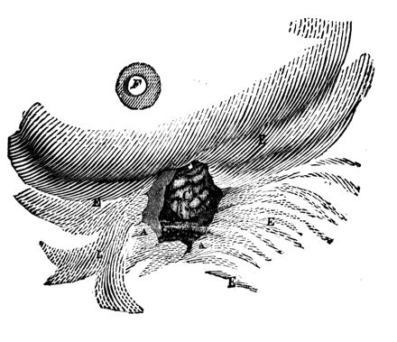
St. Martin’s recovery ran a long, tumultuous course, involving high fever, inflammation, and more surgery. Healing was eventually complete, but St. Martin was left with a gastric fistula. The stomach wound became attached to the abdomen, establishing a permanent opening from the outside of the body to the stomach cavity. A fold in the mucous lining of the stomach formed a flap inside the fistula, enabling the stomach to retain food and drink (4).
For several years, Beaumont studied the workings of St. Martin’s stomach. Beaumont observed directly the secretion of the gastric juice, and how it occurred as a response to the presence of food. He followed the digestion of various foodstuffs that St. Martin ate or Beaumont inserted into his stomach. He withdrew and examined samples of the gastric contents at intervals during the digestive process. He described the motions (contractions) of both the longitudinal and transverse muscles of the stomach, and how the pylorus allows only the chyme to pass into the duodenum.
Original drawing of St. Martin’s wound. A. Wound edges. B. Stomach cavity. C. Flap covering exposed stomach opening. E. Scar tissue surrounding the wound. F. Chest: nipple and areola.
Although isolated from the great centers of science, Beaumont corresponded with and sent samples to recognized authorities in chemistry and physiology. He published his first observations as early as January 1825, later taking St. Martin to New York to be seen by prominent physicians. Finally, in 1833 he published Experiments and Observations on the Gastric Juice and the Physiology of Digestion, a 280-page octavo volume in which he detailed his experiments, listed the inferences drawn from his data, and set forth his opinions on the subject of digestion (2). All his research, including paying St. Martin’s wages and publication of the book, was done at Beaumont’s own expense.
Sweetening the Stomach
As early as the 19th century it was recognized that in peptic ulcers (the collective term for ulcers of the stomach and duodenum) exposure of the involved tissue to acid and pepsin is essential to the development of clinical symptoms (5). Treatment accordingly was based on a two-pronged plan: 1) to lessen the amount of acid in the stomach, and 2) to coat the ulcer, minimizing further irritation by gastric juice and allowing the ulcer to heal.
Antacids
In the 20th century’s first two decades, the regimen of Dr. Bertram W. Sippy of Chicago exemplified this approach. To provide a soothing coating, Dr. Sippy made his patients drink, every hour (on the hour) from 7am to 7pm, three ounces of a mixture of equal parts milk and cream. To reduce gastric acidity, he administered an antacid powder every hour on the half hour (midway between the dairy feedings). He prescribed two different powders, of the following composition:
Sippy Powder No. 1
Bismuth subcarbonate [(BiO)2CO3] 0.65g
Sodium bicarbonate [NaHCO3] 1.3g
Sippy Powder No. 2
Magnesium oxide [MgO] 0.65g
Sodium bicarbonate [NaHCO3] 0.65g
The two powders were to be taken alternately so that Powder No. 1 would counteract the laxative effect of the magnesium oxide in Powder No. 2. Later, concern over toxicity led to the replacement of the bismuth compound by precipitated calcium carbonate (6).
With ninety years’ hindsight, the problems with the Sippy regimen are obvious. The amount of milk fat ingested would shock any modern internist or cardiologist. Also, the pre-dominant antacid in the powders was sodium bicarbonate, which would liberate significant volumes of carbon dioxide in the stomach, causing burping and general discomfort. Bicarbonate also causes rebound secretion of acid; and it can produce systemic alkalosis, especially in patients with kidney disease. There was, therefore, a movement toward antacids free of these disadvantages.
In 1929, aluminum hydroxide [Al(OH)3], in the form of a colloidal suspension (gel), was introduced in the United States (7). Slower acting than bicarbonate, aluminum hydroxide did not release CO2 or cause alkalosis, though it did tend to cause constipation. A popular proprietary form of aluminum hydroxide was Amphojel®, so named because aluminum hydroxide is amphoteric, reacting with strong alkali as well as with acid.
Many other compounds were employed as antacids. Replacing one hydroxyl of aluminum hydroxide with glycine yielded aluminum dihydroxyaminoacetate, which was recognized as an effective antacid in New and Nonofficial Remedies (NNR), a publication of the American Medical Association (8). Also enjoying some vogue was magnesium trisilicate. Less alkaline than a hydroxide, it had weaker neutralizing power, but in reacting with acid it formed insoluble silicon dioxide, which was believed to form a protective coating over the ulcer crater. Calcium carbonate (CaCO3), another effective neutralizer, is relatively tasteless and thus lends itself to use in chewable tablets (e.g., Tums®), and, in recent years, calcium carbonate has gained an additional use as a dietary supplement, to increase calcium intake.
Along with the use of antacids, efforts were made to reduce the secretion of gastric juice by oral administration of anticholinergic agents such as belladonna alkaloids (e.g., atropine, hyoscyamine, or scopolamine) and synthetic compounds of this class (e.g., homatropine methylbromide). Besides their antisecretory action, these drugs were supposed to relieve pain by an antispasmodic action on the smooth muscle of the stomach. They were usually formulated together with antacids in combination products.
A novel approach to the neutralization of gastric acid was the use of an ion-exchange resin in place of an inorganic alkali. Being insoluble and non-absorbable, ion exchangers would act only in the stomach cavity, avoiding the laxative, constipating, or alkalosis-inducing side effects of the other antacids. Such a product, Resinat®, was marketed in the mid-1940s. Each Resinat® capsule contained 250mg of a polyamine-methylene anion exchanger in basic form. The resin exchanged its hydroxyl ions for gastric chloride, thus neutralizing gastric hyperacidity (9). Despite its purported advantages, Resinat® could not compete with Amphojel® and the other conventional antacids because the resin was much more costly, and it had less neutralizing power on a gram-for-gram basis.
Through the 1950s and 1960s, the most successful antacid product was Maalox® Suspension containing, per 10mL dose, the equivalents of 440mg Al(OH)3 and 390mg Mg(OH)2. The rationale for this combination was that the constipating tendency of the aluminum and the laxative tendency of the magnesium would cancel each other out. The product’s trade name was derived from its chemical makeup: magnesium aluminum hydroxides. Maalox® was a block-buster product when blockbusters had annual sales in the millions rather than billions. It became a household word, much as Valium® and Prozac® did later on. This may have been due as much to brilliant marketing as to therapeutic effectiveness; nevertheless, the success of this simple product was the marvel and the envy of competing drug firms. New developments, as described below, have reduced the importance of Maalox®; even its trade name has been cannibalized and applied to a product containing calcium carbonate alone.
The huge number of available antacid compounds is reflected in the Food and Drug Administration (FDA) monograph for Over-the-Counter (OTC) Antacid Products. In 1972, the FDA initiated a review of all OTC products, with the plan to establish monographs describing those “recognized among qualified experts as safe and effective” (10). In each drug category, an independent advisory panel reviewed all available data. The first monograph completed, implemented in 1974, was the one for antacid products. This monograph recognizes as safe and effective: five different forms of aluminum hydroxide; three other aluminum compounds; compounds of bismuth, calcium, and magnesium; and various other substances (11).
The monograph sets two notable requirements. The recommended dose of any product must provide at least five milliequivalents of neutralizing capacity, as measured by a standard procedure in the United States Pharmacopeia (USP). Also, in combination products, each antacid ingredient must contribute at least 25% of the product’s total neutralizing capacity. This wisely limits the active ingredients to four, and avoids a situation where product A, containing twelve antacids, claims superiority over product B, which has only ten (11).
Protectants
Two protectant products––one locally and one systemically acting––are of interest. Whereas antacids have always been available over-the-counter (OTC) at the user’s discretion, these protectants are prescription-only. The locally acting product is Carafate® (sucralfate), a basic aluminum complex of sucrose octasulfate. Administered in one-gram doses, it combines with protein to form an ulcer-adherent complex at the ulcer site. It also reduces pepsin activity and has some antacid activity as well (12). Sucralfate has undergone considerable clinical study in humans.
The systemically acting protectant, Cytotec® (misoprostol) is a synthetic analog of prostaglandin E1. At therapeutic doses Cytotec® exhibits both mucosal protective and anti-secretory properties. Together, these actions reduce the risk of gastric ulcers in patients taking nonsteroidal anti-inflammatory drugs (NSAIDs), including aspirin. NSAIDs inhibit prostaglandin synthesis in the gastric mucosa, diminishing the secretion of protective mucus and bicarbonate ion (13). The high hopes once entertained for misoprostol were dashed because of its powerful abortifacient action and the finding that the proton-pump inhibitors (PPIs) (see below) are superior to misoprostol in promoting healing of active ulcers and preventing ulcer recurrence (5).
Shutting it Off at the Source
In 1976, the objective of peptic ulcer therapy changed, from neutralizing stomach acid to preventing its secretion. The first target was histamine, the decarboxylation product of the essential amino acid histidine. Histamine activity was first detected in an extract of ergot, which potently stimulates uterine contraction. Following this lead, the pioneer pharmacologists Henry Dale and George Barger isolated histamine and determined its structure in 1910 (14). Among histamine’s many pharmacologic effects is its powerful stimulation of acid release in the stomach, which was reported as early as 1930 (15). It is known now that histamine is released from enterochromaffin-like (ECL) cells, found in close proximity to the stomach’s acid-secreting (parietal) cells located in the body and fundus of the stomach. Histamine diffuses from the ECL cells into the parietal cells, where it triggers acid secretion (5).
When the first antihistamines were discovered in the 1940s, it was anticipated that they would inhibit histamine-induced gastric secretion. Surprisingly, they showed no such effect. For twenty-five years, more antihistamines were prepared, having a wide variety of chemical structures, variously sedating and not sedating. Still, their effect on stomach histamine was negligible. Until James Black came along….

An industrial researcher at ICI in the U.K., James Black discovered Inderal® (propranolol), the first beta-adrenoceptor blocking agent or “beta-blocker”. The significance of propranolol must not be underestimated. First marketed in 1964 for angina pectoris, additional indications were quickly discovered: hypertension, myocardial infarction, migraine, and glaucoma. The field of cardiovascular diseases––previously limited to drugs like digitalis, nitroglycerin, and quinidine––was opened wide to the promise of more specific, effective drug therapy.
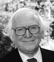
Black did not rest on his laurels. He speculated that, just as there were at least two adrenergic receptors, there might be more than one type of receptor for histamine. About this time, he changed jobs, moving to Smith Kline & French (SKF) Research Institute in Welwyn where, in 1964, Black and Michael Parsons developed a screening procedure to measure the ability of experimental compounds to prevent acid secretion in the stomach of anesthetized rats. Four years and more than 200 synthetic histamine analogs later, his group had found nothing yet. [Ed.: See also our Crosstalk interview with Sir James Black in the June 2004 issue of Molecular Interventions.]
At this point a crucial event occurred. SKF headquarters in Philadelphia instructed Black to abandon the seemingly futile project. Black and his group kept right on working, but imposed “radio silence” by not sending any further reports to the head office. There were more disappointments, including a promising compound (Metiamide), which went into clinical trial but quickly proved too toxic. Finally, in 1976, twelve years after Black’s initial experiments, Tagamet® (cimetidine) was brought to the market (16). At last the gastric acid could be cut off at its source! Cimetidine, the original H2 receptor antagonist, was soon followed by ranitidine (Zantac®), famotidine (Pepcid®), and nizatidine (Axid®).
There is still another level of complexity to gastric acid secretion. Histamine’s effect is to stimulate the cAMP-dependent pathway within the parietal cell. A second pathway, dependent on calcium ions (but independent of histamine’s actions), is activated by other physiologic agonists. Both pathways––the histamine-dependent and the calcium-dependent––culminate in the activation of the H+,K+-ATPase system (the so-called proton pump) (5). In the 1970s and 1980s, small-molecule chemical structures were discovered that could inhibit the proton pump. These PPIs are the most effective suppressors of gastric-acid secretion because they act at the very end of both biochemical pathways, thus, the PPIs block gastric acid (i.e., proton) secretion in both the histamine-dependent and the histamine-independent (calcium-dependent) pathways. The PPIs approved in the United States are omeprazole (Prilosec®), lansoprazole (Prevacid®), rabeprazole (Aciphex®), and pantoprazole (Protonix®). Thus, the intimate knowledge of cell physiology, receptors, and the biochemistry of the stomach developed in the final decades of the twentieth century revolutionized the treatment of peptic ulcers and related conditions [e.g., gastro-esophageal reflux disease (GERD) and Zollinger-Ellison syndrome].
A New Germ Is Discovered
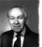
In the early 1980s, while the R&D laboratories of “big pharma” were racing to discover new H2 blockers and PPIs that would not infringe another firm’s patents, a young medical resident, Barry Marshall, and a pathologist, Robin Warren, working in Perth, Western Australia, were making a revolutionary observation. Their discovery was made possible by a technological advance of the previous decade-the perfection of the endoscope, which allowed gastroenterologists to look directly at stomach tissue, perform biopsies, and diagnose gastritis and peptic ulcer more accurately (17). Marshall and Warren examined biopsy specimens from the antral mucosa of 100 patients undergoing endoscopy for various ailments. In fifty-eight of these patients they found spiral or curved bacteria. The bacteria were present in nearly all patients with chronic active gastritis, gastric ulcer, or duodenal ulcer (18). They were flagellate, best seen by staining with silver, and slow growing in culture. At first thought to be a new species of Campylobacter, they were later recognized as a new genus and named Helicobacter pylori.
In their original report Marshall and Warren (18) noted that, since 1940, spiral gastric bacteria had several times been observed, reported and forgotten. Indeed, Sippy in 1915 (6) mentioned earlier work by E.C. Rosenow blaming a streptococcus for weakening the stomach lining. It was probably easy to “forget” these reports because it seemed inconceivable that bacteria could live at the pH of gastric juice. The stomach is the human’s first line of defense against microorganisms in his food.
Thus, even the very existence of H. pylori flew in the face of conventional scientific wisdom. Even harder to stomach––so to speak––was Marshall’s insistence that this bacterium was causally associated with gastritis and peptic ulcers, conditions long believed to be largely stress-induced. Add to this his lack of established research credentials and the fact that, like William Beaumont, he came from “nowhere,” and it is easy to understand the establishment’s skepticism. Unable to produce ulcers in laboratory animals injected with H. pylori, Marshall was driven to offer himself as guinea pig. Early one morning in July, 1984 Marshall went to his laboratory and, without informing either his wife or the hospital’s ethics board of his plan, swallowed a live culture of the organisms (17).
The outcome of the experiment was fortunate for Marshall. After a one-week latent period, he was sick for about a week with headaches, foul-smelling breath, and achlorhydria. He became pale, tired, and hoarse. During that week he underwent endoscopy, which showed his stomach lining to be inflamed, and a biopsy that revealed swarms of bacteria around inflamed stomach cells. Several days later, all clinical symptoms had ceased and a repeat biopsy showed that everything had returned to normal; Marshall’s body cleared the infection without medication. Marshall had made his case and researchers worldwide began studying H. pylori.
The presence of H. pylori has been demonstrated in a sizable portion of the general population. The theory now is that its spiral shape and flagella enable it to penetrate the mucous layer of the stomach wall, where it is shielded from the gastric acid (19). H. pylori is now accepted as a major cause of chronic type-B gastritis, which is closely associated with peptic ulcer (19). Medical opinion generally holds that eradicating this organism prevents the recurrence of ulcers once they are healed.
H. pylori is sensitive to many antibiotics, and also to the non-antibiotic bismuth subsalicylate and bismuth subcitrate; however, use of a single agent does not produce a permanent result. For permanent clearance, the so-called triple therapy must be used consisting of a bismuth compound plus two antibiotics (chosen from amoxicillin, clarithromycin, metronidazole, and tetracycline). Addition of a PPI is recommended, especially when the acid-sensitive antibiotics amoxicillin or clarithromycin are used (5). Serendipitously, bismuth subsalicylate happens to be the active ingredient of Pepto-Bismol®, an OTC preparation marketed for many years for upset stomach and diarrhea.
A Century of Progress
For seventy-five years, treatment of peptic ulcer and related gastric maladies was based mainly on antacids, with an ongoing pursuit of products having improved side effect profiles and palatability. In the century’s final quarter, remarkable new developments practically tumbled over each other. These began with systemically acting drugs to reduce the secretion of gastric acid––first the H2 receptor antagonists (e.g., cimetidine), followed by the more efficient proton pump inhibitors (e.g., omeprazole). Finally, the discovery of H. pylori forced a rethinking of long-established concepts as to the etiology of peptic ulcers and the best mode of treatment.
Progress has sprung from basic research in biochemistry and cell metabolism, and from advances in technology of medical instrumentation. Recognition is also due to three major figures: William Beaumont, who supported his research from his own salary; Sir James Black, who risked his position by flouting the corporate hierarchy; and Barry Marshall, who gambled his very health to prove his hypothesis.
- © American Society for Pharmacology and Experimental Theraputics 2005

Stanley Scheindlin, DSc, RPh, holds graduate degrees in pharmaceutical chemistry and worked in drug product development and regulatory affairs. Now retired, he is a part-time consultant and writes freelance articles for pharmacy-related specialty publications.


