|
 
 |
| ORIGINAL ARTICLE |
|
| Year : 2016 | Volume
: 4
| Issue : 1 | Page : 1-7 |
|
Relationship between age at menarche and anthropometric profiles, demography, and ethnicity among adolescent girls in Nigeria
Monday Nwankwo, Barnabas Danborno, Wilson Oliver Hamman
Department of Human Anatomy, Ahmadu Bello University, Zaria, Nigeria
| Date of Web Publication | 13-Sep-2016 |
Correspondence Address:
Monday Nwankwo
Department of Human Anatomy, Faculty of Medicine, Ahmadu Bello University, Zaria
Nigeria
 Source of Support: None, Conflict of Interest: None  | Check |
DOI: 10.4103/2315-7992.190468

Introduction: This study was made to evaluate and compare the age at menarche among Nigerian schoolgirls from Hausa and Igbo ethnic groups, and to examine whether ethnicity, demographics, and selected anthropometric characteristics of the studied population influence age at menarche. This was a retrospective cohort study. Materials and Methods: Data pertaining to menarche, ethnicity, and demographics were collected using questionnaires from 800 secondary schoolgirls of Nigeria, in 2014. Six hundred (600) were postmenarcheal, while 200 were premenarcheal. Among the schoolgirls, 300 postmenarcheal and 100 premenarcheal girls were of Hausa and Igbo descent, respectively. We compared the mean age at menarche of girls based on ethnicity, demographics, and the anthropometric parameters of menstruating and nonmenstruating girls. The relationship between anthropometric indices and age at menarche was determined. Results: Mean age at menarche among girls from Igbo ethnicity is lower than that of girls of Hausa ethnicity. Compared to rural girls, girls from urban centers reach menarche earlier. The mean age at menarche of the entire sample population was 13.54 ± 0.90 years. Mean ages at menarche of Hausa and Igbo girls were 13.65 ± 0.92 years and 13.44 ± 0.87 years, respectively. Mean body mass index (BMI) values in postmenarcheal and premenarcheal age were 20.50 ± 2.13 kg/m2 and 17.64 ± 1.40 kg/m2, respectively. The mean weight and height of menstruating girls were 49.02 ± 3.85 kg and 154.92 ± 0.05 cm, respectively, while those of nonmenstruating girls were 38.83 ± 2.60 kg and 147.56 ± 3.54 cm, respectively. Conclusion: Ethnicity, demographics, and selected anthropometric measurements have influence on age at menarche. Girls of Igbo ethnicity reach maturity earlier than those of Hausa descent, girls in urban centers have lower age at menarche, and the anthropometric measurements of menstruating girls are higher than those of nonmenstruating girls. Keywords: Adolescent girls, anthropometry, demographics, ethnicity, menarche
How to cite this article:
Nwankwo M, Danborno B, Hamman WO. Relationship between age at menarche and anthropometric profiles, demography, and ethnicity among adolescent girls in Nigeria. Ann Bioanthropol 2016;4:1-7 |
How to cite this URL:
Nwankwo M, Danborno B, Hamman WO. Relationship between age at menarche and anthropometric profiles, demography, and ethnicity among adolescent girls in Nigeria. Ann Bioanthropol [serial online] 2016 [cited 2017 Jan 3];4:1-7. Available from: http://www.bioanthrojournal.org/text.asp?2016/4/1/1/190468 |
| Introduction | |  |
Menarche is a distinct maturational event in female childhood. It represents the hallmark of the physical events occurring in the hypothalamic-pituitary-gonadal axis. During fetal, neonatal, and early infancy periods, the gonadotropin-releasing
hormone (GnRH) is active. Pulsatile secretion of GnRH at puberty and the episodic secretion of pituitary gonadotropin hormone for normal gonadal development and function are stimulated by the activation of the GnRH pulse generator.[1] The secretion of GnRH helps in the regulation of growth, development, and function of the ovaries in females by signaling the pituitary gland to secrete luteinizing hormone (LH) and follicle-stimulating hormone (FSH), otherwise known as gonadotropins. In females, the gonadotropins play significant roles in ovulation (release of an ovum from a mature Graafian follicle). FSH stimulates the development and maturation of a follicle in one of the ovaries in females.
This paper will discuss the influence of demographics in determining the menacheal age among two ethnic groups, the Hausa and the Igbo in Nigeria. Several studies from various countries worldwide have shown that menarcheal age is influenced by various factors including genetics, ethnicity, nutrition, socioeconomic status, physical activity, body mass index (BMI), and demographics.[2],[3],[4],[5],[6],[7] Padez [8] documented a downward trend in age of menarche among the Portuguese and the influence of place of residence in childhood on menarche.
The effect of urbanization on menarcheal age has also been widely reported: That urban girls mature earlier, on average, than rural girls.[9],[10],[11],[12],[13],[14],[15] The decline in menarcheal age began in the late 19th century in Europe.[16] Socioeconomic status, often measured by parental educational attainment or occupation, has also been hypothesized to influence menarcheal age—girls from families with higher socioeconomic class tend to reach menarche earlier than those from families with lower socioeconomic status. This is seen in the findings of Bielicki et al.[17] in Poland; Attallah et al.[18] in Sudan; and Oduntan et al.[15] and Uche and Okorafor [14] in Nigeria. A spectrum of factors other than those mentioned before that have significant influence on menarcheal age include family size and birth order, with girls from larger-sized families reaching menarche later than those from smaller-sized families.[19],[20],[21] Nutrition [22] has also been reported to have influence on menarcheal age.
Family size and birth order currently seem to have relatively consistent influence, irrespective of country, on menarcheal age. By the same token, parents' educational background and occupation seem to have a weak correlation with menarcheal age in industrialized countries, due to the homogenous nature of such societies.[19],[23],[24] On the other hand, the heterogeneous nature of developing countries may be accountable for the significant influence parents' educational attainment and occupation has on menarcheal age.
Risk factors associated with early menarche include type 2 diabetes,[25],[26] breast cancer,[27],[28] and adolescent risk-taking behaviors.[29] Further, early menarche has also been associated with anxiety symptoms, depression, early sexual initiation, and cardiovascular incidents.[1]
The aim of the present study was to assess mean age at menarche in girls of two ethnic groups in Nigeria (schoolgirls of Hausa and Igbo descent), and to assess the influence of geographical location at childhood and adolescence on the menarcheal age of Nigerian secondary schoolgirls. We also wanted to investigate the difference in anthropometric measurements, particularly BMI, in menstruating and nonmenstruating Nigerian secondary schoolgirls.
| Materials and Methods | | |
The target population in our sample was selected randomly from among secondary schoolgirls of the participating schools in 2014. Data were obtained from the participants using predesigned questionnaires, after which their anthropometric measurements were taken. Girls with any form of abnormality that may affect their normal growth or endocrine system were excluded from the study. The study protocol was reviewed and approved by the Health Research Ethics Committee of the Ahmadu Bello University Teaching Hospital, Zaria, Nigeria. Consent to participate in the study was obtained from the relevant authorities of participating schools and from the participants themselves. Only those who gave informed consent to participate were included in the study.
Study subjects
A total of 800 girls aged 11-18 years participated in the present cross-sectional study, of whom 400 were of Hausa ethnicity, while the remaining 400 were of Igbo descent. Of the 400 from each ethnicity, 200 lived in the rural area, while the remaining 200 were urbanites from childhood (lasts to age 7) to adolescence (older than age 7).[30] One hundred fifty (150) of the 200 girls living in rural area and 150 of the 200 girls living in the urban area were postmenarcheal, while the remaining 50 (each) were premenarcheal.
Anthropometric measurements
Anthropometric measurements on participants were carried out by a well-trained female research assistant according to the following protocols. Height was measured to the nearest centimeter using a portable stadiometer. Weight was recorded without shoes and wearing lightweight clothes to 0.1 kg. BMI was calculated with height and weight [weight (kg)/height (m 2)]. Chest circumference was recorded at the level of the middle of the sternum (breastbone), with the tape passing under the arms to the nearest 0.1 cm. After the tape was in position, the arms were to be relaxed by the side, and the measurement was taken at the end of a normal expiration. Hip circumference was recorded with the tape wrapped over the largest part of the buttocks to the nearest 0.1 cm. Mid-upper arm circumference was measured by asking the participant to flex the bicep muscle while the tape was wrapped around the flexed bicep, halfway between the shoulder and the elbow, also to the nearest 0.1 cm. Thigh circumference was measured at the level of the mid-point on the lateral (outer side) surface of the thigh, midway between the trochanterion (top of the thigh bone or femur) and the tibia lateral (top of the tibia bone). Waist circumference measurements were taken to the nearest 0.1 cm by measuring from the narrowest point between the lower borders of the rib cage and the iliac crest at the end of a normal expiration. The current age and menarcheal age of each girl were asked. Place of residence at childhood and adolescence were noted and classified into two categories: Rural centers and urban centers.
Statistical analyses
Data were analyzed in SPSS version 20 (IBM Corporation, Zaria and Nsukka, New York, USA). P values smaller than 0.05 (two-tailed) were considered statistically significant. The following statistical tests were carried out: Mean and standard deviation (SD), Student's t-test, analysis of variance (ANOVA), Pearson correlation, and linear regression.
| Results | | |
The sample size of this experiment was 800, and 200 of the girls had not already had their first menses at the time of this study. Thus, the mean age of menarche was calculated for 600 postmenarcheal girls and for the 200 premenarcheal girls who participated in the exercise.
The mean age of menarche for all girls sampled was 13.46 ± 0.90 years [Table 1]. There were significant differences between the mean ages of menarche for Hausa (13.65 ± 0.92 years) and the mean ages of menarche for Igbo (13.44 ± 0.87 years) girls, t = -2.96, P < 0.01. There was a significant difference between the mean anthropometric measures for Hausa and the mean anthropometric measures for Igbo girls, with the girls from Igbo ethnic group scoring on the average higher than their counterparts from Hausa ethnicity, P < 0.05 [Table 1].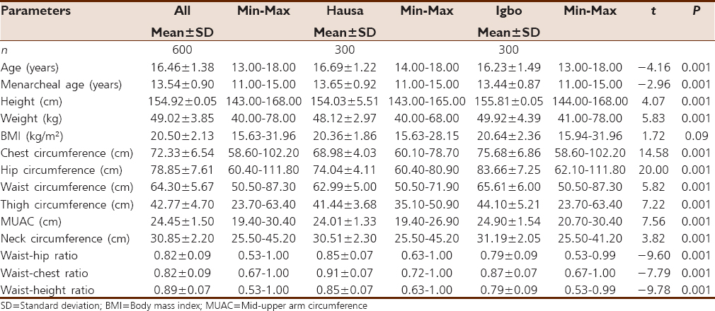 | Table 1: Mean and SD of menarcheal age and body composition of all subjects and for subjects according to ethnicity
Click here to view |
[Table 2] shows the results of comparison of menarcheal age and anthropometric measurements based on place of residence at childhood in girls from the two ethnic groups. From the table, girls from rural areas irrespective of ethnicity were found to tend to have a delay in their maturation compared to those residing in urban areas, with a mean age at menarche of 13.83 ± 0.95 years and 13.48 ± 0.86 years for girls of Hausa descent and 13.52 ± 0.87 years and 13.35 ± 0.88 years for girls of Igbo ethnicity (P < 0.05 and P > 0.05), respectively. Considering girls of Hausa ethnicity, there is a statistically significant difference in the mean anthropometric measurements based on geographical location, with girls from the urban environment scoring on the average higher than girls from rural places (P < 0.05) except for BMI, waist circumference, and neck circumference. These exceptional measures did not show statistical significance (P > 0.05). On the other hand, anthropometric measurements of girls of Igbo ethnicity indicated that geographical location has low effect on these measurements, with hip circumference, waist circumference, mid-upper arm circumference, and neck circumference showing statistical significance (P < 0.05). | Table 2: Mean and SD of menarcheal age and body composition of Hausa and Igbo subjects from rural and urban areas
Click here to view |
Ethnicity and place of residence at childhood are associated with being older or younger at menarche. [Table 3] compares the age at menarche of girls from rural areas of girls from Hausa and Igbo ethnic groups; the same comparison was done for girls from urban areas. From the table, it is obvious that girls of Igbo ethnicity reach menarche earlier for comparisons done for rural and urban places. This is a higher-bound estimate, as it is not just based on current residence. The higher figure observed for girls residing in rural areas is related to a number of variables, such as poverty, malnutrition, educational attainment, and poor sanitary and health conditions. Result in [Table 4] shows comparison of menarcheal age based on nutritional status of the girls. From the table, thin subjects reach menarche later than their overweight counterparts. This difference is statistically significant P < 0.001. There was an inverse correlation between BMI and age at menarche [Table 5]. Though age at menarche had a statistically significant negative correlation with BMI, weight, hip circumference, and chest circumference, it had, however, a positive correlation with waist-hip ratio and waist-height ratio. No statistically significant association was found between age at menarche and height, waist circumference, mid-upper arm circumference, neck circumference, thigh circumference, and waist chest ratio, with P > 0.05.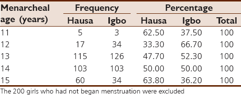 | Table 3: Frequency and percentages of menarcheal ages of subjects according to ethnicity
Click here to view |
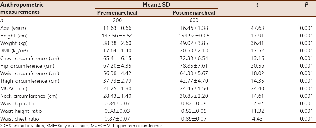 | Table 5: Comparison of mean anthropometric variables between menstruating and nonmenstruating females
Click here to view |
[Table 6] shows the regression analyses equations to analyze which were the most predictive variables. Weight and BMI were the variables selected for predicting age at menarche (P ≤ 0.001). [Table 7] presents predictive equations for estimating menarcheal age of subjects based on ethnicity.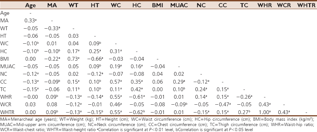 | Table 6: Correlation matrix of menarcheal age and anthropometric characteristics of girls from both ethnic groups (n=600)
Click here to view |
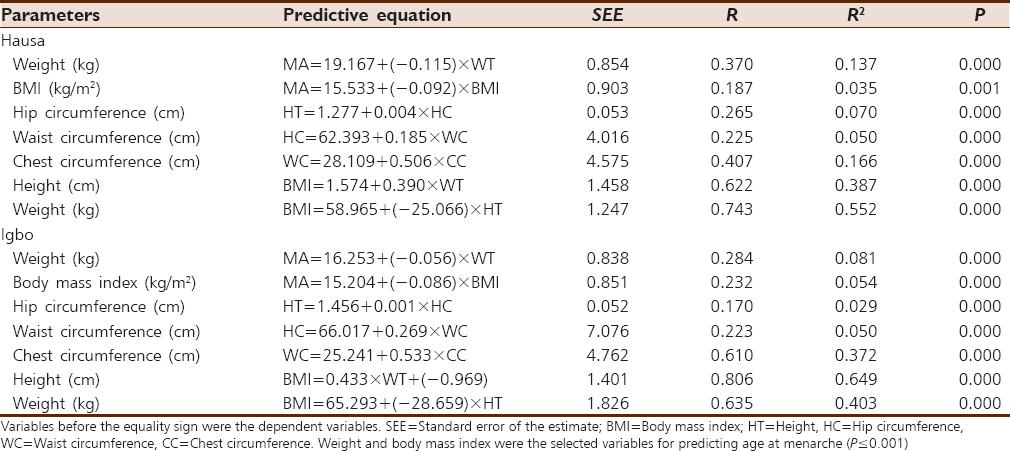 | Table 7: Multiple regression analyses of anthropometric variables of girls from Hausa and Igbo ethnic groups
Click here to view |
| Discussion | | |
In this group of contemporary girls, we have found a number of associations between ethnicity, geographical location, some anthropometric parameters, and the occurrence of menarche. The mean age of menarche for our study cohort was 13.54 ± 0.90 years and the age of onset of menarche in the majority of our subjects, or 84.33% (not shown), was 13-14 years. Our findings are important because of their potential impact on early-matured girls' behaviors.
Demography, ethnicity, and menarche
Girls in urban areas reach maturity at earlier ages than those from a rural environment. The trend in age of menarche due to place of residence at childhood found in our study was similar to that reported by Uche and Okorafor,[14] Oduntan et al.[15] in Nigeria, Pasquet et al.[10] in Cameroon, and Marrodan et al.[31] in Spain. The urban-rural variations may be due to the fact that various urban centers tend to have much better health-care facilities, better sanitary standards of dwellings, and better schools, among several other characteristics.[17]
Other studies support our results indirectly. Abubakar et al.[32] in a study of 381 schoolgirls from the Hausa ethnic group obtained a mean age at menarche of 13.67 ± 1.20 years, while Chukwujekwu et al.[33] in 2014 reported a mean age of menarche for 341 secondary schoolgirls from the Igbo ethnic group to be 13.49 ± 0.06 years. These two studies were conducted in urban areas. According to Hughes et al., genes are responsible in determining the rate of developmental maturation and age at menarche for about 75% of the difference in girls from the same ethnic group, monozygotic twins, and mother/daughter pairs. Talma et al. 2013[2] found a lower median age at menarche among Turkish and Moroccan girls as compared to Dutch girls.
BMI, weight, height, and menarche
The link between obesity and early maturity among girls has been reported by several studies.[34],[35] Early menarche is suggested to be associated to obesity. This is, however, controversial, as early age at menarche has also been found among girls with normal weight.[36] Obesity is thought to influence menarche by preventing pregnancy until there is enough adiposity to sustain the conceptus and the mother.[37] The mean values of anthropometric parameters were significantly higher among menstruating girls when compared to nonmenstruating ones. The present results showed a negative correlation between BMI and age at menarche; this is congruous with the report of Pierce and Leon.[38] Results from the present study also revealed the sensitivity of BMI percentile to nutritional status and the onset of menarche. Differences in BMI between premenarcheal and postmenarcheal girls have previously been reported by several authors.[5],[37],[39],[40] At less than fifth percentile of BMI, only 28 girls were menstruating with age at menarche of 13.79 ± 0.83 years, while those predisposed to overweight with BMI greater than the 85th percentile (68 girls) were menstruating at an early age of 13.16 ± 1.01 years.
In spite of the fact that the mechanisms involved in the negative relationship between obesity and age at menarche are not well understood, hormonal factors are alleged to influence the rate of puberty through fat accumulation in the body, as noted by Van et al.[41] A minimum weight of 48.80 kg is required for the onset of menstruation,[42] with body adiposity accounting for 23.7%. In our study, the minimum weight for menstruating girls was 40 kg, while the average weight values for postmenstrual and premenarcheal girls were 49.02 ± 3.85 kg and 38.38 ± 2.60 kg, respectively. This is in agreement with several other studies.[43],[44],[45]
The issue of influence of height on age at menarche is somewhat controversial. There was a time when menarcheal age was thought to be dependent on height.[6] The secular trend seems to have stopped in developed countries.[4],[46],[47],[48] In our study, there is a weak correlation between height and early onset of menarche. Hence, increase in height may not be responsible for the decline in age at menarche. However,[49] and Kaplowitz et al.[50] showed that taller girls reach maturation earlier than shorter ones. It has been reported that tall but heavier girls reached menarche earlier than short, slender ones.[51] Early puberty among females results in early closure of the epiphyseal plates due to estrogenic secretion from the ovary, which leads to low chances of increase in height after puberty, as discussed by Onland-Moret et al.[52] Such females record a slight increase in height after puberty.
| Acknowledgments | | |
We would like to express our sincere appreciation to the study participants for their patience, cooperation, and participation. The authors are also grateful to the various school authorities for providing permission and invaluable contribution that led to the successful conduction of the study. The help of our research assistants is also very much appreciated.
| References | | |
| 1. | Talma H, Schoönbeck Y, van Dommelen P, Bakker B, van Buuren S, Hirasing RA. Trends in menarcheal age between 1955 and 2009 in the Netherlands. PLoS One 2013;8:e60056.  |
| 2. | Karapanou O, Papadimitriou A. Determinants of menarche. Reprod Biol Endocrinol 2010;8:115. |
| 3. | Aksglaede L, Juul A, Olsen LW, Sorensen TI. Age at puberty and the emerging obesity epidemic. PLoS One 2009;4:e8450. |
| 4. | Gohlke B, Woelfle J. Growth and puberty in German children: Is there still a positive secular trend? Dtsch Arztebl Int 2009;106:377-82. |
| 5. | Anderson SE, Must A. Interpreting the continued decline in the average age at menarche: Results from two nationally representative surveys of U.S. girls studied 10 years apart. J Pediatr 2005;147:753-60. |
| 6. | Mul D, Fredriks AM, van Buuren S, Oostdijk W, Verloove-Vanhorick SP, Wit JM. Pubertal development in The Netherlands 1965-1997. Pediatr Res 2001;50:479-86. |
| 7. | Fredriks AM, van Buuren S, Burgmeijer RJ, Meulmeester JF, Beuker RJ, Brugman E, et al. Continuing positive secular growth change in The Netherlands 1955-1997. Pediatr Res 2000;47:316-23. |
| 8. | Padez C. Social background and age of menarche in Portuguese university students: A note on the secular changes in Portugal. Am J Hum Biol 2003;15:415-27. |
| 9. | Graham MJ, Larsen U, Xu X. Secular trend in age at menarche in China: A case study of two rural counties in Anhui Province. J Biosoc Sci 1999;31:257-67. |
| 10. | Pasquet P, Biyong AM, Rikong-Adie H, Befidi-Mengue R, Garba MT, Froment A. Age at menarche and urbanization in Cameroon: Current status and secular trends. Ann Hum Biol 1999;26:89-97. |
| 11. | Barnes-Josiah D, Augustin A. Secular trend in the age at menarche in Haiti. Am J Hum Biol 1995;7:357-62. |
| 12. | Hulanicka B, Waliszko A. Deceleration of age at menarche in Poland. Ann Hum Biol 1991;18:507-13. |
| 13. | Cameron N, Wright CA. The start of breast development and age at menarche in South African black females. S Afr Med J 1990;78:536-9. |
| 14. | Uche GO, Okorafor AE. The age of menarche in Nigerian urban school girls. Ann Hum Biol 1979;6:395-8. |
| 15. | Oduntan SO, Ayeni O, Kale OO. The age of menarche in Nigerian girls. Ann Hum Biol 1976;3:269-74. |
| 16. | Ahmed ML, Ong KK, Dunger DB. Childhood obesity and the timing of puberty. Trends Endocrinol Metab 2009;20:237-42. |
| 17. | Bielicki T, Szklarska A. Secular trends in stature in Poland: National and social class-specific. Ann Hum Biol 1999;26:251-8. |
| 18. | Attallah NL. Age at menarche of schoolgirls in Egypt. Ann Hum Biol 1978;5:185-9. |
| 19. | Roberts DF, Rozner LM, Swan AV. Age at menarche, physique and environment in industrial North East England. Acta Paediatr Scand 1971;60:158-64. |
| 20. | Billewicz WZ, Fellowes HM, Thomson AM. Menarche in Newcastle upon Tyne girls. Ann Hum Biol 1981;8:313-20. |
| 21. | Apraiz AG. Influence of family size and birth order on menarcheal age of girls from Bilbao City (Biscay, Basque Country). Am J Hum Biol 1999;11:779-83. |
| 22. | Simondon KB, Simon I, Simondon F. Nutritional status and age at menarche of Senegalese adolescents. Ann Hum Biol 1997;24:521-32. |
| 23. | Demoulin F. Secular trend in France. In: Bodzsa'r, E. Susanne, C. editors. Secular Growth Changes in Europe. Budapest: Eotvos University Press; 1998. p. 109-34. |
| 24. | Dann TC, Roberts DF. Menarcheal age in university of Warwick young women. J Biosoc Sci 1993;25:531-8. |
| 25. | Stöckl D, Döring A, Peters A, Thorand B, Heier M, Huth C, et al. Age at menarche is associated with prediabetes and diabetes in women (aged 32-81 years) from the general population: The KORA F4 Study. Diabetologia 2012;55:681-8. |
| 26. | He C, Zhang C, Hunter DJ, Hankinson SE, Buck Louis GM, Hediger ML, et al. Age at menarche and risk of type 2 diabetes: Results from 2 large prospective cohort studies. Am J Epidemiol 2010;171:334-44. |
| 27. | He C, Chasman DI, Dreyfus J, Hwang SJ, Ruiter R, Sanna S, et al. Reproductive aging-associated common genetic variants and the risk of breast cancer. Breast Cancer Res 2012;14:R54. |
| 28. | Velie EM, Nechuta S, Osuch JR. Lifetime reproductive and anthropometric risk factors for breast cancer in postmenopausal women. Breast Dis 2005-2006;24:17-35. |
| 29. | Michaud PA, Suris JC, Deppen A. Gender-related psychological and behavioural correlates of pubertal timing in a national sample of Swiss adolescents. Mol Cell Endocrinol 2006;254-255:172-8. |
| 30. | Bogin B. Patterns of Human Growth. 2 nd ed. Cambridge, UK: Cambridge University Press; 1999. |
| 31. | Marrodán MD, Mesa MS, Aréchiga J, Pérez-Magdaleno A. Trend in menarcheal age in Spain: Rural and urban comparison during a recent period. Ann Hum Biol 2000;27:313-9. |
| 32. | 32. Panti AA, Ekele BA, Nwobodo EI, Ahmed Y. The age at menarche amongst secondary school girls in Sokoto metropolis, North-West Nigeria. Open J Microphysics 2011;23:1-4. |
| 33. | Chukwujekwu IE, Ezejindu DN, Olabisi OJ. Age, Weight, Height, Body Mass Index and Waist-Circumference at Menarche among Secondary School Girls in Otolo, Nnewi, South Eastern Nigeria. Int J Res 2014;1:4. |
| 34. | Roede MJ, van Wieringen JC. Growth diagrams 1980: Netherlands third nation-wide survey. TSG 1985;63(Suppl):1-34. |
| 35. | Cole TJ, Bellizzi MC, Flegal KM, Dietz WH. Establishing a standard definition for child overweight and obesity worldwide: International survey. BMJ2000;320:1240-3. |
| 36. | Aksglaede L, Sørensen K, Petersen JH, Skakkebaek NE, Juul A. Recent decline in age at breast development: The Copenhagen puberty study. Pediatrics 2009;123:e932-9. |
| 37. | Kaplowitz PB. Link between body fat and the timing of puberty. Pediatrics 2008;121(Suppl 3):S208-17. |
| 38. | Pierce MB, Leon DA. Age at menarche and adult BMI in the Aberdeen children of the 1950s cohort study. Am J Clin Nutr 2005;82:733-9. |
| 39. | Anderson SE, Dallal GE, Must A. Relative weight and race influence average age at menarche: Results from two nationally representative surveys of US girls studied 25 years apart. Pediatrics 2003;111:844-50. |
| 40. | O'Dea J, Abraham S. Should body-mass index be used in young adolescents? Lancet 1995;345:657. |
| 41. | van Lenthe FJ, Kemper CG, van Mechelen W. Rapid maturation in adolescence results in greater obesity in adulthood: The Amsterdam Growth and Health Study. Am J Clin Nutr 1996;64:18-24. |
| 42. | Frisch RE, Revelle R. Height and weight at menarche and a hypothesis of menarche. Arch Dis Child 1971;46:695-701. |
| 43. | Goon DT, Toriola AL, Uever J, Wuam S, Toriola OM. Growth status and menarcheal age among adolescent school girls in Wannune, Benue State, Nigeria. BMC Pediatr 2010;10:60. |
| 44. | Kim JY, Oh IH, Lee EY, Oh CM, Choi KS, Choe BK, et al. The relation of menarcheal age to anthropometric profiles in Korean Girls. J Korean Med Sci 2010;25:1405-10. |
| 45. | Khakbazan Z, Niroomanesh SH, Mehran A, Majidi-Ahi A. To investigate the menarcheal age and it, relation with body mass index. Iranian J Hayat 2005;11:55-62. |
| 46. | Schönbeck Y, Talma H, van Dommelen P, Bakker B, Buitendijk SE, HiraSing RA, et al. The world's tallest nation has stopped growing taller: The height of Dutch children 1955 to 2009. Pediatr Res 2013;73:371-7. |
| 47. | Freedman DS, Khan LK, Serdula MK, Srinivasan SR, Berenson GS. Secular trends in height among children during 2 decades: The Bogalusa Heart Study. Arch Pediatr Adolesc Med 2000;154:155-61. |
| 48. | Brundtland GH, Liestøl K, Walløe L. Height, weight and menarcheal age of Oslo schoolchildren during the last 60 years. Ann Hum Biol 1980;7:307-22. |
| 49. | Kaplowitz P. Pubertal development in girls: Secular trends. Curr Opin Obstet Gynecol 2006;18:487-91. |
| 50. | Akhavan-Karbasi S, Bafghi M. Determination of beginning of puberty age and its factors on Yazd girl students. Iranian J Shahid Sadooghi Yazd Medical Sciences University 1997;5:26. |
| 51. | Kaplowitz PB, Slora EJ, Wasserman RC, Pedlow SE, Herman-Giddens ME. Earlier onset of puberty in girls: Relation to increased body mass index and race. Pediatrics 2001;108:347-53. |
| 52. | Onland-Moret NC, Peeters PH, van Gils CH, Clavel-Chapelon F, Key T, Tjønneland A, et al. Age at menarche in relation to adult height: The EPIC study. Am J Epidemiol 2005;162:623-32. |
[Table 1], [Table 2], [Table 3], [Table 4], [Table 5], [Table 6], [Table 7]
|


 Search Pubmed for