|
 
 |
| ORIGINAL ARTICLE |
|
| Year : 2016 | Volume
: 4
| Issue : 1 | Page : 60-63 |
|
A study of the near and far interpupillary distances among the Bura ethnic group of North-Eastern Nigeria
Yohanna Musa Usman, Ali Ishaq Shugaba
Department of Human Anatomy, Faculty of Medical Sciences, University of Jos, Jos, Nigeria
| Date of Web Publication | 13-Sep-2016 |
Correspondence Address:
Yohanna Musa Usman
Department of Human Anatomy, Faculty of Medical Sciences, University of Jos, Jos
Nigeria
 Source of Support: None, Conflict of Interest: None  | Check |
DOI: 10.4103/2315-7992.190465

Introduction: The aims of this study were to establish standard for the interpupillary distance, document the anthropometric variation of this parameter with advancing age and determine the extent of sexual dimorphism of this parameter among the Bura ethnic group of North Eastern Nigeria. Materials and Methods: 300 subjects comprising of 150 males and 150 females of Bura ethnic group with ages ranging from 7 to 40 were recruited for this study. Data were analyzed in statistical software (SPSS for Windows, Version 17.0, Chicago: Inc.) and comparative tests were conducted using the independent student's -t-test at significant level of 0.05. Results: The mean Near Interpupillary Distance was 63 ± 6.2 mm while the mean Far Interpupillary distance was 69 ± 6.4 mm. The mean values for the Near and Far Interpupillary Distances were 63.2 ± 6.5 mm and 69.6 ± 6.7 mm for males and 62.6 ± 5.9 mm and 68.0 ± 5.9 mm for females respectively. Conclusion: In conclusion, these findings would be of benefit in the diagnosis of craniofacial syndromes associated with hyper/hypotelorism, management of posttraumatic orbitofacial deformities, the manufacture of spectacle frames and lenses and as a guide for dentists in selecting denture teeth. Keywords: Bura, far interpupillary distance, near interpupillary distance, North-Eastern Nigeria
How to cite this article:
Usman YM, Shugaba AI. A study of the near and far interpupillary distances among the Bura ethnic group of North-Eastern Nigeria. Ann Bioanthropol 2016;4:60-3 |
| Introduction | |  |
Anthropology is the scientific and humanistic study of the human species. It is the exploration of the human diversity in time and space.[1] Anthropometry is the measurement of human body parts and dimensions.[1] It is divided into four parts: Osteometry, cephalometry, somatometry, and craniometry.[1] Anthropometry is done on living people as well as on skeletal remains from sites.[1]
Interpupillary distance (IPD) has been defined by various authors as the distance between the centers of the pupils.[2] It has been revealed that IPD is the best indicator of the distance between the centers of the two eye globes.[2] The clinical observation of the face, especially the orbital region, remains an essential part for the clinical evaluation of phenotypic anomalies, which can be either qualitative or quantitative anomalies. Diagnosis of quantitative anomalies as hypertelorism requires a knowledge of the normal variation of the trait in a population of a given ethnic background and at a certain age.[3]
The mean IPD depends on the characteristics of the population from which the data are drawn. It is statistically significantly different between the two genders, between certain racial/ethnic groups, between near and far viewing, and between certain age groups.[1],[4] Mean and median IPD for the adult population appear to lie somewhere near 63 mm.[1],[4] With regard to far IPD (FIPD), the vast majority of adults lie within the range of 50–75 mm. There are several cases of people outside this range and there is at least one case of a 15-year-old female with an IPD of 43 mm.[1],[4]
It has been reported that the near IPD (NIPD) and the FIPD for males are significantly larger than those of females in the Ijaw people (P< 0.001) and that the NIPD in the Ijaws at 8–12 years was 57.13 ± 3.23 mm and 56.01 ± 2.92 mm for males and females, respectively. The overall mean values for FIPD obtained for the same population were 59 ± 4.4 mm and 59 ± 5.2 mm for males and females, respectively.[5]
Reported studies have shown that FIPD for male and females was 7.18 cm and 7.10 cm, respectively, and that in the 7–15-year-old girls in the Igbo ethnic group, the values for FIPD and NIPD were 6.47 cm and 5.9 cm, respectively.[6]
The overall FIPD obtained in a Turkish population was 60.75 ± 4.03 mm for men and boys and 59.45 ± 3.51 mm for girls and women. In the 7–11 year age group, the average IPD was greater in the Turkish population (54.5–59 mm)[7] than the reported averages for Chinese (52 mm), Negro (53.1–57.5 mm), and Caucasian children (52–56 mm).[7] The Turkish values are very similar to those reported for Hong Kong (54–59 mm) and British children (55–60 mm).[7] In the 7–15-year-old girls, Turkish FIPD and NIPD values were 58.03 ± 3.31 and 55.31 ± 3.29 mm, respectively, which are quite similar with the values of Arabian children, which are 57.55 ± 3.29 and 55.32 ± 3.29 mm, respectively.[7]
The mean IPD of the subjects selected from the Department of Prosthodontics, Lahore Medical and Dental College, Lahore, was shown to be 65.26 ± 5.41 mm by Hussain et al. with a range of 81.29 mm to 44.41 mm.[8] However, other studies showed a mean value of 59.16 mm after measuring 100 subjects of the United States army, 63.51 mm for 109 edentulous patients with a range of 38.00–73.00 mm. Hussain et al.[8] also revealed an increased IPD values for males than females, and these gender-based variations similar to Hussain et al.'s study were also reported in other studies.[8]
The aims of this study were to establish a standard for the IPD, document the anthropometric variation of this parameter in different age groups, and determine the extent of sexual dimorphism of this parameter among the Bura ethnic group of North-Eastern Nigeria. These findings would be of benefit in the diagnosis of craniofacial syndromes associated with hyper/hypotelorism, management of post-traumatic orbitofacial deformities, the manufacture of spectacle frames and lenses, and as a guide for dentists in selecting denture teeth.
| Materials and Methods | | |
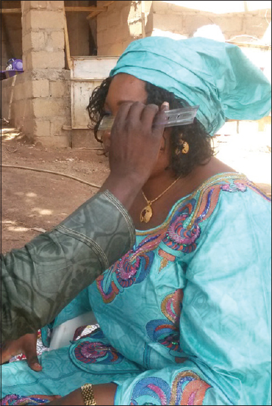
This was a cross-sectional study conducted among the Bura ethnic group of North-Eastern Nigeria, in which 300 subjects comprising 150 males and 150 females with ages ranging from 7 to 40 were recruited. The lower limit of the age range was based on the fact that younger age group may not cooperate with the examiner whereas the upper limit of age was based on the assumption that any change in the measured parameters would have stopped by the age of 40 years. Ethical clearance and informed consents were obtained. Subjects were seated comfortably in a chair and the examiner also sat in front of them with the subject's and examiner's heads at the same level. Measurement of the IPD was performed with a nonstretchable plastic ruler [Figure 1]. The millimeter ruler was held tightly against the subject's nasal bridge. The examiner closed his right eye first and asked the subject to look at the examiner's opened left eye. The 0 mark on the ruler was placed at the outer (temporal) limbus margin of the subject's right eye while the examiner sighted with his open left eye, the point on the ruler that corresponded to the inner (nasal) limbus of the subject's left eye. This measurement is equivalent to the NIPD. The examiner then closed his left eye and asked the subject to look at the examiner's open right eye. Still maintaining the 0 mark on the ruler at the outer (temporal) limbus of the subject's right eye, the examiner sighted the point on the ruler that corresponded to the inner (nasal) limbus of the subject's left eye. This measurement is equivalent to FIPD. The same procedure was repeated from right to left for every subject and recorded.
The data were entered and analyzed in statistical software (SPSS for Windows, Version 17.0, Chicago: Inc). The comparative tests were conducted using the independent Student's t-test at a significant level of 0.05 to demonstrate any statistical significance between the measured parameters.[9]
| Results | | |
The results of this study are presented in [Table 1],[Table 2],[Table 3],[Table 4],[Table 5]. Subjects were divided into three age groups; 7–15 years, 16–25 years, and 26–40 years to determine the anthropometric variation of the measured parameters to determine the relationship of the measured parameters with various age groups. The mean values in the text are represented as mean value ± standard deviation.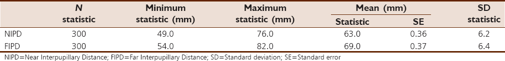 | Table 1: Overall mean values of near interpupillary distance and far interpupillary distance
Click here to view |
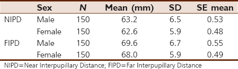 | Table 2: Mean values of near interpupillary distance and far interpupillary distance with respect to sex
Click here to view |
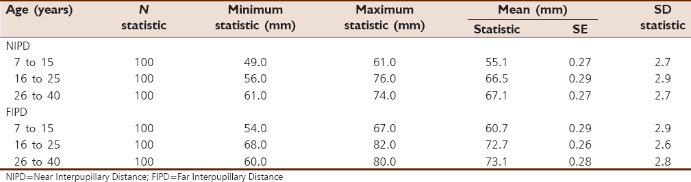 | Table 3: Mean values of near interpupillary distance and far interpupillary distance with respect to age
Click here to view |
 | Table 4: Anthropometric variation of near interpupillary distance and far interpupillary distance with age
Click here to view |
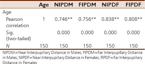
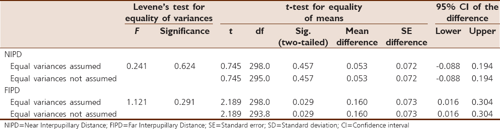 | Table 5: Sexual dimorphism of near interpupillary distance and far interpupillary distance
Click here to view |
The mean NIPD of the subjects in this study was 63 ± 6.2 mm [Table 1]. The mean values for the NIPD and FIPD were 63.2 ± 6.5 mm and 69.6 ± 6.7 mm for males and 62.6 ± 5.9 mm and 68.0 ± 5.9 mm for females respectively [Table 2].
For the age groups 7–15, 16–25, and 26–40 years, the mean values for NIPD were 55.1 ± 2.7 mm; 66.5 ± 2.9 mm; and 67.1 ± 2.7 mm, respectively [Table 3]. There is a strong correlation between different age groups and the mean values of NIPD and FIPD in all age groups and among both sexes signifying anthropometric variation with age [Table 4]. There is a demonstration of significant sexual dimorphism among NIPD and FIPD [Table 5].
| Discussion | | |
Ocular dimensions are important in the diagnosis and treatment of congenital orbital or craniofacial anomalies and post-traumatic deformities as well as in the proper mounting of spectacle lenses to eliminate unwanted prismatic effect.[10] Normal values of IPD and intercanthal distance are important for the successful reconstruction of the canthal area. Thus, it is important to have local data of these parameters since this standard reflect the potentially different pattern of craniofacial growth resulting from racial, ethnic, sexual, and age differences.[10]
This study has revealed that the overall mean NIPD and FIPD were 63 ± 3.6 mm and 69 ± 3.7 mm, respectively [Table 1], and were, respectively, 63.2 ± 5.3 mm and 69.6 ± 5.5 mm for males and 62.6 ± 4.8 mm and 68.0 ± 4.9 mm for females [Table 2]. These values are lower than those reported for the Igbos by Esomonu et al.[6] who had a mean FIPD of 71.8 mm and 71.0 mm for males and females, respectively. The Buras in this study, however, show higher mean values than the Ijaws who were reported to have FIPD of 59 ± 4.4 mm and 59 ± 5.2 mm for males and females, respectively, by Osunwoke et al.[5] The mean values reported for the Tusks and those reported by Hussain et al. were similar to those of the Buras in this study, though slightly lower.[8]
This study shows a strong correlation between different age groups and the mean values of NIPD and FIPD in all age groups and among both sexes signifying anthropometric variation with age of all parameters [Table 3] and [Table 4]. This is in agreement with a study among the Igbo ethnic group, which reported higher values of FIPD in the older age group of that study.[6]
It has been demonstrated in this study that there is a significant sexual dimorphism among the mean values of NIPD and FIPD [Table 5]. This is in agreement with the study done by Osunwoke et al.[5] among the Ijaw ethnic group which revealed higher mean values of NIPD and FIPD in males than in females. Esomonu et al. also reported higher mean values in males than in females of both NIPD and FIPD.[6] Indeed, several other studies have also demonstrated a significant sexual dimorphism in NIPD and FIPD.[7], 8, [10],[11],[12],[13]
| Conclusion | | |
This study has established standards for the IPD in the Bura ethnic group of North-Eastern Nigeria, demonstrated the anthropometric variation of the studied parameters with age, and showed that there is a significant sexual dimorphism with higher mean values in males than in females. These findings would be of benefit in the diagnosis of craniofacial syndromes associated with hyper/hypotelorism and management of post-traumatic cranial and orbitofacial deformities in the manufacture of spectacle frames and lenses and as a guide for dentists in selecting denture teeth.
Declaration of patient consent
The authors certify that they have obtained all appropriate patient consent forms. In the form the patient(s) has/have given his/her/their consent for his/her/their images and other clinical information to be reported in the journal. The patients understand that their names and initials will not be published and due efforts will be made to conceal their identity, but anonymity cannot be guaranteed.
Financial support and sponsorship
Nil.
Conflicts of interest
There are no conflicts of interest.
| References | | |
| 1. | Conrad PK. Anthropology: The Exploration of Human Diversity. 10 th ed. London, United Kingdom: University of Michigan, McGraw Hill; 2004. p. 3-77.  |
| 2. | Pryor HB. Objective measurement of interpupillary distance. Pediatrics 1969;44:973-7. |
| 3. | Dollfus H, Verloes A. Dysmorphology and the orbital region: A practical clinical approach. Surv Ophthalmol 2004;49:547-61. |
| 4. | Evereklioglu C, Doganay S, Hamdi ER, Gunduz A. Distant and near interpupillary distance in 3448 and female subjects: Final results. Turgut Ozal Tip Merkezi Dergisi 1999;6:84-91. |
| 5. | Osunwoke EA, Didia BC, Olotu EJ, Yerikema AH. A study on the normal values of inner canthal, outer canthal, interpupillary distance and head circumference of 3-12 years Ijaws. Am J Sci Ind Res 2012;3:441-5. |
| 6. | Esomonu UG, Taura MG, Anas IY, Madibbo MH. Anthropometric studies of the interpupillary distance among the Igbos of the South Eastern Nigeria. Bajopas 2012;5:123-6. |
| 7. | Evereklioglu C, Doganay S, Er H, Gunduz A, Tercan M, Balat A, et al. Craniofacial anthropometry in a Turkish population. Cleft Palate Craniofac J 2002;39:208-18. |
| 8. | Hussain MW, Qamar K, Naeem S. The role of interpupillary distance in the selection of anterior teeth. Pak Oral Dent J 2012;32:165-9. |
| 9. | Daniel WW. Biostatistics. A Foundation for Analysis in the Health Science. USA: John Wiley and Sons; 1987. p. 207-18. |
| 10. | Oladipo GS, Okoh PD, Hart JS. Anthropometric study of ocular dimensions in adult ijaws of Nigeria. Res J Med Med Sci 2010;5:121-4. |
| 11. | Charles OA, Hakeem FB, Nervey DW, Mildred BA. Normal outer and inner canthal measurements of the ijaws of Southern Nigeria. Eur J Sci Res 2008;22:163-7. |
| 12. | Oladipo GS, Akande PA, Osogba IG, Yorkum KL. Anthropometric studies of inner canthal distance, outer canthal distance and canthal index of adult ibibios. Asian J Med Sci 2011;3:14-6. |
| 13. | Etezad-Razavi M, Jalalifar S. Correlation between interpupillary and Inner – Outer intercanthal distances in individuals younger the 20. J Ophthalmic Vision Res 2008;3:16-22. |
[Figure 1]
[Table 1], [Table 2], [Table 3], [Table 4], [Table 5]
|


 Search Pubmed for