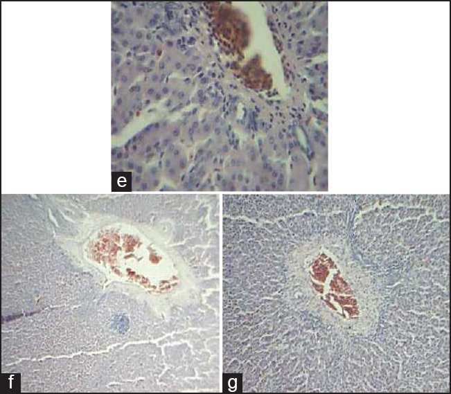
Figure 2: Comparative histology illustrations of the bird. (e) Section of the liver of Gallus gallus domesticus domesticus showing a portal triad (pt) composed of a large vein, artery and bile ductile H&E × 400 (f) Section of the liver of Gallus gallus domesticus domesticus showing a portal triad (pt) composed of a large vein, artery and bile ductile H&E × 40 (g) Section of the liver of Gallus gallus domesticus domesticus indicating central vein H&E × 100