Close
  Indian J Med Microbiol
 

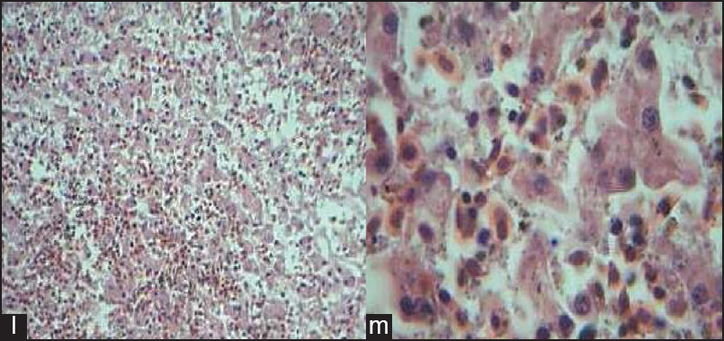
Figure 4: Comparative histology illustrations of the amphibian liver. (l) Section of the liver of Rana tigrinas showing hepatocytes disposed in singles and cluster, separated by a loose fibrovascular connective tissue stroma. Nucleated red cells are within the intervening sinusoids H&E × 100 (m) Section of the liver of Rana tigrinas showing hepatocytes disposed in singles and cluster, separated by a loose fibrovascular connective tissue stroma. Nucleatedred cells are within the intervening sinusoids H&E × 400

Figure 4: Comparative histology illustrations of the amphibian liver. (l) Section of the liver of <i>Rana tigrinas</i> showing hepatocytes disposed in singles and cluster, separated by a loose fibrovascular connective tissue stroma. Nucleated red cells are within the intervening sinusoids H&E × 100 (m) Section of the liver of <i>Rana tigrinas</i> showing hepatocytes disposed in singles and cluster, separated by a loose fibrovascular connective tissue stroma. Nucleatedred cells are within the intervening sinusoids H&E × 400