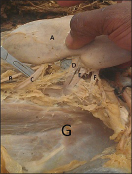
Figure 2: The posterior surface of the left kidney as it is pulled away from the paravertebral gutter, together with the structures entering and leaving the substance of the kidney (A: Left kidney; B: Left ureter; C: First accessory renal artery; D: Main renal artery; E: Second accessory renal artery; F: Third accessory renal artery; G: Left paravertebral gutter)