|
 
 |
| CASE REPORT |
|
| Year : 2014 | Volume
: 2
| Issue : 3 | Page : 86-88 |
|
Ocular Prosthesis.-A unique method, for post evisceration ocular defect
Kalpana Devaraju, Harish Gopalkrishna, Sanjana Rao
Dayananda Sagar College of Dental Sciences and Hospital, Kumaraswamy Layout, Bangalore, Karnataka, India
| Date of Web Publication | 10-Sep-2014 |
Correspondence Address:
Harish Gopalkrishna
Department of Prosthdontics, Dayananda Sagar College of Dental Sciences and Hospital, kumaraswamy layout, Type 2, No 7, 1st cross, 7th main, BTM 2nd stage Bangalore - 560 076, Karnataka
India
 Source of Support: None, Conflict of Interest: None  | Check |
DOI: 10.4103/2347-4610.140517

Ocular prosthesis is artificial replacement of the eye, while orbital prosthesis is replacement of entire content of the orbit. Replacement of the missing eye is not only important but also has to be immediate, as the psychological trauma associated with it is overcome better. This case presents a unique way of replacing the missing eye, where-in a combination of prefabricated eye and impression for customizing eye prosthesis was adopted.
Keywords: Ocular prosthesis, orbital prosthesis, prefabricated eye
How to cite this article:
Devaraju K, Gopalkrishna H, Rao S. Ocular Prosthesis.-A unique method, for post evisceration ocular defect
. Eur J Prosthodont 2014;2:86-8 |
| Introduction | |  |
Eye is not only the most important sense organ, but also plays an important role in esthetic appearance and facial expression. Eye or eyes may be missing due to congenital anomaly, trauma, or pathology which necessitates its surgical removal. Peyman, Saunders, and Goldberg have classified surgical removal of eyes into three types as evisceration where contents of the globe are removed leaving the sclera intact, enucleation where the entire eyeball is removed after severing the muscles and optic nerve, and exenteration where contents of the orbit including the eyelids and surrounding tissues are removed. [1],[2]
Replacement of missing eye has to be done immediately due to as the psychological trauma associated with it, is overcome better and there is improved social acceptance. Ocular prosthesis is artificial replacement of the bulb of the eye, while orbital prosthesis is replacement of the entire contents of orbit. [3] In the case presented here, a unique way of fabricating eye prosthesis was followed for evisceration defect.
| Case Report | | |
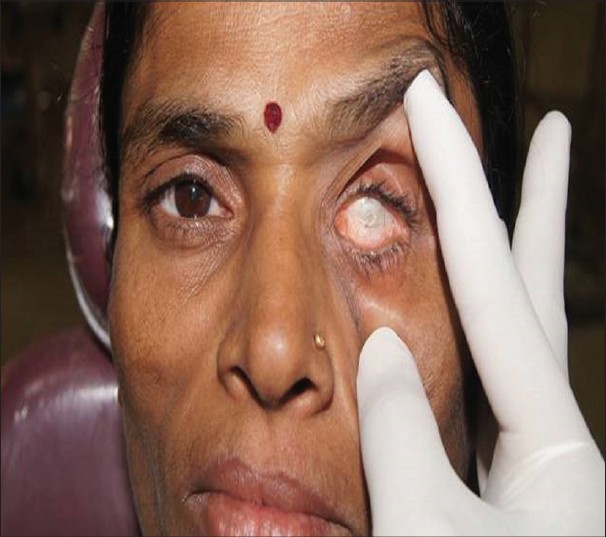
A 54-year-old female patient reported to the Department of Prosthodontics, Dayananda Sagar College of Dental Sciences and Hospital, Bangalore, with the chief complaint of missing left eye and wanted replacement to be done.
On investigation, the patient gave a history of surgical removal due to glaucoma. Examination revealed evisceration of the eye with healthy intraocular tissue bed. There was sufficient depth between upper and lower fornices for retention of ocular prosthesis [Figure 1].
Iris button of the stock eye was used for fabricating the eye prosthesis to the patient. Even though conventional technique for creating an iris button is esthetically better, it is more technique sensitive and laborious. Hence, stock eye was used for the present case.
Conformer was used as tray for making impression of the ocular defect. It was first tried by placing in the defect and the necessary over-extensions were trimmed. Petroleum jelly was applied to the eyelashes. Disposable syringe was connected to the conformer. Light body additional silicone (Aquasil-; Dentsply detry, Gmbh, 78467 Konstanz, Germany) was mixed and dispensed into the syringe. Conformer was placed into the defect and light body was injected. Patient was asked to perform all the movements of the eye to record the exact depth and width of the defect [Figure 2].
After the material set, the entire assembly was removed from the defect. Putty consistency of the additional silicon (Aquasil) was used to completely cover the impression to create a mold space [Figure 3].
White wax was poured into the mold after removal of the impression. The wax model trial was done and necessary corrections were made [Figure 4].
Prefabricated eye shell which matched with the iris of contralateral eye was selected. Iris button of the eye shell was trimmed. It was made 0.5 mm short all around than the iris of the natural eye, to compensate for the magnification caused by a layer of heat-cure acrylic in the definitive prosthesis.
Iris button was placed over the wax model after removing a layer of wax from the wax model. The positioning of iris button was replicated as that in the contralateral eye [Figure 5].
Two-piece flasks were used for processing, wherein the iris button was in one portion of the flask and the remaining part in the other portion. Tooth-colored heat-cure acrylic (Dental Product of India) of exact shade was used for replacing sclera portion of the eye prosthesis. Veining was added by attaching red-dacron fibers to the prosthesis using the monomer-polymer syrup. Then, 0.5-1 mm of acrylic was trimmed from the frontal portion of acrylized prosthesis. Again, heat-cure acrylic was added on to the frontal portion and processing was completed. The prosthesis was retrieved, finished, polished, and delivered to the patient.
Patient was given instructions regarding the usage and maintenance of prosthesis. Recall visit was done and necessary corrections were made [Figure 6].
| Discussion | | |
Presently, three types of ocular prosthesis are in use,: stock eyes, stock eyes modified by various methods, and custom-fitted eyes. [4] The materials used range from glass to methyl methacrylate to silicones. Glass eye is difficult to manufacture, hazardous, [5] heavy and brittle. Methyl methacrylates have superior strength, and permit modifications in shape and size. [5] Silicones, being flexible, are advantageous when in contact with movable tissue beds. [5]
In the present case, the prosthesis was fabricated using a combination of iris button from prefabricated eye and customized scleral portion from impression of the socket. In ocular prosthesis, positioning of iris button becomes important for better esthetics. [6] Ocular impressions and fitting can be placed into one of several broad categories as follows: direct impression/external impression, impression with stock ocular tray or modified stock ocular tray, impression with custom ocular tray, impression using stock ocular prosthesis, ocular prosthesis modification, and wax scleral blank technique. [7]
In this method, the disadvantages of prefrabricated eye, such as poor fit and movement of the prosthesis, were overcome and the cumbersome steps involving painting of iris button for custom eye prosthesis were eliminated. However, limitatations of the technique were availability of prefabricated eye with perfect match of iris button, color stability of heat-cure resins, and polymerization process of amide-perioxide system which progresses more slowly and is less complete than barbituric acid initiator system. [8],[9]
Definitive prosthesis should have the following characteristics: [10]
- Retain the shape of the defective socket
- Prevent collapse or loss of the shape of the lids
- Provide proper muscular action of the lids
- Prevent accumulation of fluid in the cavity
- Maintain palpebral opening similar to the natural eye
- Mimic the coloration and properties of the natural eye
- Has gaze similar to the natural eye.
| References | | |
| 1. | Perman KI, Baylis HI. Evisceration, enucleation, and exenteration. Otolaryngol Clin North Am 1988;21:171-82. 
|
| 2. | Agrawal KK, Mall P, Alvi H A, Rao J, Singh K. Fabrication of custom made prosthesis for anophthalmic paediatric patients: 2 case reports. J Interdicp Dent 2012;2:128-31.
|
| 3. | Kumar CH, Sajjan CS. Prosthetic management of an ocular defect. Contemp Clin Dent 2010;1:201-3.
|
| 4. | Jayaswal GP, Dange SP, Khalikar AN. Restoration of an atrophic eye socket with custom made eye prosthesis, utilizing digital photography. Indian J Dent Res 2011;22:482-5.
[PUBMED]  |
| 5. | Kaira LS, Bhayana R, Asopa V, Pandey AN, Dabral E. Prosthetic management of ocular defect: A case series. Eur J Prosthodont 2014;2:33-6.
|
| 6. | Guttal SS, Patil NP, Vernekar N, Porwal A. A simple method of positioning the iris disk on a custom made ocular prosthesis. A clinical report. J Prosthodont 2008;17:223-7.
|
| 7. | Mathews MF, Smith RM, Sutton AJ, Hudson R. The Ocular Impression: A Review of the Literature and Presentation of an Alternate Technique. J Prosthodont 2000;9:210-6.
|
| 8. | Kale E, Mese A, Izgi AD. A Technique for Fabrication of an Interim Ocular Prosthesis. J Prosthodont 2008;17:654-61.
|
| 9. | Scheuermann H. Residual monomer content in prosthetic materials. Dent Labor (Munch) 1981;29:1695-6.
|
| 10. | Haug SP, Andres CJ. Fabrication of custom ocular prosthesis. In: Taylor TD, editor. Clinical maxillofacial prosthetics. 1 st ed. Chicago: Quintessence Publishing; 2000. p. 265-76.
|
[Figure 1], [Figure 2], [Figure 3], [Figure 4], [Figure 5], [Figure 6]
|


 Search Pubmed for