|
 
 |
|
IMAGE |
|
|
|
| Year : 2011 | Volume
: 17
| Issue : 1 | Page : 33-34 |
| |
A case of short stature with anterior vertebral beaking
Ketan Prasad Kulkarni, Inusha Panigrahi
Genetic-Metabolic Unit, Department of Pediatrics, Postgraduate Institute of Medical Education and Research (PGIMER), Chandigarh, India
| Date of Web Publication | 18-Jun-2011 |
Correspondence Address:
Inusha Panigrahi
Pediatric Genetics, Advanced Pediatric Center, PGIMER, Chandigarh -12
India
 Source of Support: None, Conflict of Interest: None
DOI: 10.4103/0971-6866.82191

How to cite this article:
Kulkarni KP, Panigrahi I. A case of short stature with anterior vertebral beaking. Indian J Hum Genet 2011;17:33-4 |
How to cite this URL:
Kulkarni KP, Panigrahi I. A case of short stature with anterior vertebral beaking. Indian J Hum Genet [serial online] 2011 [cited 2016 May 13];17:33-4. Available from: http://www.ijhg.com/text.asp?2011/17/1/33/82191 |
A 20-month-old female child from Chandigarh, India, presented with poor gain in length since birth, short limbs, and dysmorphic facies. The child was a product of a nonconsanguineous marriage and was born to a 22-year-old father and 20-year-old primigravida mother by lower segment caesarian section (indication: premature rupture of membranes) at 32 weeks of gestation. There was no history of drug intake in early gestation or of antenatal radiation exposure. On examination, the length, weight, and occipitofrontal circumference of the child were 60.6 cm (<3 standard deviation), 6.7 kg (<3 standard deviation), and 44.8 cm (<3 standard deviation), respectively. The upper segment to lower segment ratio was 2.1: 1. The child had triangular facies with brachycephaly. He had acromesomelic shortening of limbs with short stubby fingers. There was no kyphoscoliosis, organomegaly, or polydactyly. Neurological examination was within normal limits with no developmental delay. There was no cherry red spot on fundus examination. The hemogram, serum calcium, phosphate and alkaline phosphate, liver function tests, and renal function tests were within normal limits. Skiagram of the spine (lateral view) showed anterior beaking of vertebrae [Figure 1]. X-ray of wrist with hand revealed short metacarpals, phalanges, and conical middle phalanges [Figure 2]. The rest of the skeletal survey was within normal limits. The urine analysis for mucopolysaccharides was twice negative.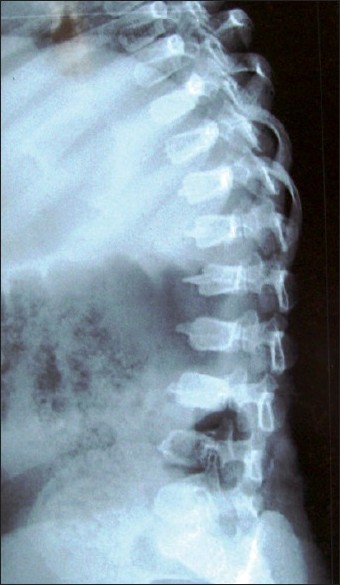 | Figure 1: Skiagram of the spine (lateral view) showing platyspondyly, pear-shaped vertebrae with anterior beaking of T12 to L4 vertebrae
Click here to view |
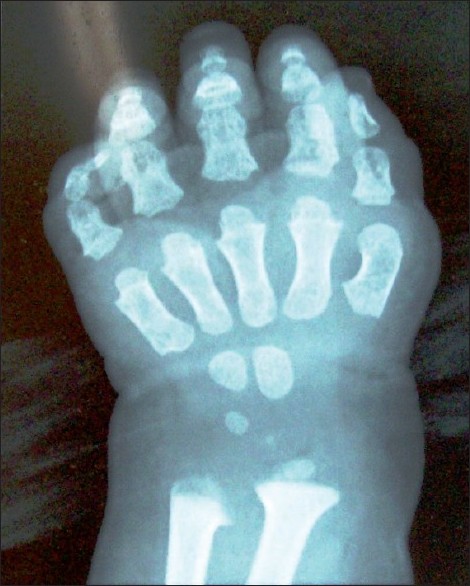 | Figure 2: X-ray of wrist with hand revealed irregular ulnar and radial metaphysis, short tubular metacarpals, short phalanges, and conical distally tapering middle phalanges.
Click here to view |
In view of the striking anterior beaking of the vertebrae, the initial diagnosis considered in the index patient was Morquio syndrome, but the radiological features in the skiagram of the wrist and hand were inconsistent with the diagnosis. [1] Similarly, urine analysis was not supportive. On further elucidation, the clinical presentation, investigations, and radiological features were found to be consistent with 'spondylometaepiphyseal dysplasia, short limb-hand type' (SEMD). [1],.[2],[3] Patients with this clinical entity usually have normal intellectual development. In view of its severe and life-threatening complications like spinal cord damage due to atlantoaxial instability and its variable clinical spectrum, pediatricians, radiologists, and geneticists must be aware of this nosologic entity. [1] Classical radiological features in SEMD can guide appropriate investigations and diagnosis, given the paucity of literature available on this nosologic entity. Prenatal diagnosis and genetic counseling may be offered in select cases.
 References References | |  |
| 1. | Online Mendelian Inheritance in Man. OMIM (TM). Mckusick-Nathans Institute for Genetic Medicine, John Hopkins University (Baltimore, MD) and National Center for Biotechnology Information, National Library of Medicine (Bethesda, MD), accessed September 2008. 
|
| 2. | Borochowitz Z, Langer LO Jr, Gruber HE, Lachman R, Katznelson MB, Rimoin DL. Spondylo-meta-epiphyseal dysplasia (SMED), short limb-hand type: A congenital familial skeletal dysplasia with distinctive features and histopathology. Am J Med Genet 1993;45:320-6.
[PUBMED] |
| 3. | Lakhar BN, Raphael R. Spondyloepiphyseal dysplasia: An evaluation of six cases. Indian J Radiol Imaging 2003;13:199-203.
 |
[Figure 1], [Figure 2]
|


