|
 
 |
|
CASE REPORT |
|
|
|
| Year : 2013 | Volume
: 19
| Issue : 1 | Page : 87-89 |
| |
Tracheal agenesis with broncho-esophageal fistula in VACTERL / TACRD association
Suresh R. S. Mandrekar, Sangeeta Amoncar, R. G. W Pinto
Department of Pathology, Goa Medical College, Bambolim, Goa, India
| Date of Web Publication | 4-Jun-2013 |
Correspondence Address:
Suresh R. S. Mandrekar
2 Antil Peth, Bicholim - 403 504, Goa
India
 Source of Support: None, Conflict of Interest: None
DOI: 10.4103/0971-6866.112910

 Abstract Abstract | | |
Tracheal agenesis (TA) is an extremely rare malformation. We report here autopsy findings in a case of TA with bronchoesophageal fistula of Floyd type III. The other malformations present included laryngeal atresia, Right lung hypolobulation, ventricular septal defect in membranous portion, bilateral cystic renal dysplasia, spleninculus, Meckel's diverticulum, and imperforate anus. The constellations of malformations present in our case have overlapping features with Vertebral anomalies, Anal atresia, Cardiovascular anomalies, Tracheo-esophageal fistula, Esophageal atresia, Renal anomalies, Limb anomalies and Tracheal atresia or laryngo tracheal atresia, Cardiac anomalies, Renal anomalies, Duodenal atresia association described previously in the literature.
Keywords: Tracheal atresia or laryngo tracheal atresia, Cardiac anomalies, Renal anomalies, Duodenal atresia association, Tracheal agenesis, Vertebral anomalies, Anal atresia, Cardiovascular anomalies, Tracheo-esophageal fistula, Esophageal atresia, Renal anomalies, Limb anomalies association
How to cite this article:
Mandrekar SR, Amoncar S, Pinto R. Tracheal agenesis with broncho-esophageal fistula in VACTERL / TACRD association. Indian J Hum Genet 2013;19:87-9 |
How to cite this URL:
Mandrekar SR, Amoncar S, Pinto R. Tracheal agenesis with broncho-esophageal fistula in VACTERL / TACRD association. Indian J Hum Genet [serial online] 2013 [cited 2016 May 24];19:87-9. Available from: http://www.ijhg.com/text.asp?2013/19/1/87/112910 |
| Introduction | |  |
Tracheal agenesis (TA) is a rare congenital anomaly, which results inevitably in immediate respiratory distress after delivery. Since, the first report of the case in 1900 [1] approximately, 150 cases have been reported in the world literature. TA is classified by Floyd et al.[2] into three categories depending upon the fistulous communication of the trachea/bronchi with the esophagus (broncho-esophageal fistula). TA seldom occurs as an isolated anomaly and is most often associated with multisystem congenital malformations. Various malformations seen with TA form patterns, which overlap with Vertebral anomalies, Anal atresia, Cardiovascular anomalies, Tracheo-esophageal fistula, Esophageal atresia, Renal anomalies, Limb anomalies association but are distinct and is considered to be a part of another association called Tracheal atresia or laryngo tracheal atresia, Cardiac anomalies, Renal anomalies, Duodenal atresia. [3],[4]
| Case Report | | |
A pre-term baby (30 weeks) that was small for gestational age (weight 1.370 Kg) delivered by spontaneous vaginal delivery by a unbooked primigravida mother was admitted in neonatal ICU with respiratory distress syndrome and died within 2 h of birth. The ante-mortem diagnosis was tracheo-esophageal cleft with respiratory distress with imperforate anus. A post mortem examination was performed to determine the type of tracheo-esophageal abnormality. At autopsy there was cyanosis and imperforate anus. There were no other external congenital abnormalities.
On dissection of the upper airways the epiglottis was normal and there was complete laryngeal atresia. On bisecting through the atretic segment the laryngeal cartilages were well formed [Figure 1]. The esophagus was opening normally in the hypopharynx. In the lower part of the esophagus openings of both the main bronchi were seen separately [Figure 2] and [Figure 3] forming broncho-esophageal fistula.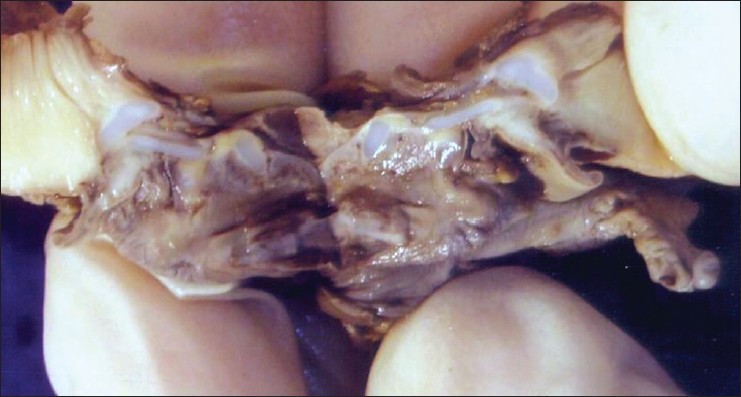 | Figure 1: Gross bisected specimen of the larynx showing atresia of lumen with normal cartilages
Click here to view |
 | Figure 2: En block gross specimen showing the lumen of the esophagus with depressions of the bronchial openings
Click here to view |
The left lung was grossly normal whereas the right lung showed no separation into distinct lobes (hypolobulation). Both the lungs appeared airless and solid. Microscopic examination revealed alveoli of both the lungs to be filled with fibrinous material and hemorrhage.
The esophagus was continuous with stomach, which was normal. There was no hiatus or diaphragmatic hernia. The duodenum was normal and there was no stenosis or atresia. A Meckel's diverticulum was present in the ileum. A spleninculus was present.
Both the kidneys were grossly cystic with atretic ureters [Figure 4]. Microscopically both the kidneys revealed cystic renal dysplasia [Figure 5]. The heart showed a ventricular septal defect in the membranous portion and the cardiac chambers, valves, and great vessels were normal. | Figure 5: Photomicrograph showing cartilage in cystic renal dysplasia (H and E, ×400)
Click here to view |
| Discussion | | |
Congenital malformations affecting the trachea and oesophagus are not infrequent; however, the certain forms are quite rare. TA is an extremely rare anomaly with only about 150 cases reported in the world literature.
The classification of TA by Floyd et al.[2] is the most universally accepted. They classified TA into three variants. In Type I, a short segment of distal trachea arises from the anterior wall of the esophagus before dividing into the main stem bronchi. In Type II, there is complete agenesis of the trachea with a fistula between the esophagus and carina from which the two main-stem bronchi originate. In Type III, the two main stem bronchi arise individually from the anterior esophageal wall. The relative incidence of the three types is 13%, 65%, and 22% respectively and the present case fits into TA Floyd's type III.
TA of all the three types seldom occur as an isolated anomaly and most of the reports in the literature mention about multiple associated abnormalities. [5]
The present case had laryngeal atresia, right lung hypolobulation, ventricular septal defect, cystic renal dysplasia, spleninculus, Meckel's diverticulum, and imperforate anus.
The association of TA with other multiorgan congenital malformations has been a topic of interest. The high incidence of associated anomalies has led some authors to suggest that TA may be one of the components of VACTERL association. [6] VACTERL association includes Vertebral anomalies, Anal atresia, Cardiovascular anomalies, Tracheo-esophageal fistula, esophageal atresia, renal anomalies and pre-axial limb anomalies. Three of the seven defects are considered to be sufficient to include the case as VACTERL association. Laryngo-tracheo-esophageal clefts, complete laryngeal agenesis, and webbing of the vocal cords have also been described in association with TA. [7]
Using these criteria, the present case can be considered as a part of VACTERL association.
Some authors however, believe that TA is not a part of VACTERL association but is one of the malformation in a different pattern of association known as TACRD that includes TA or laryngo-tracheal atresia, complex Congenital cardiac anomalies, Renal anomalies, and Duodenal atresia. [3],[4] The acronym TACRD covers most of the common congenital anomalies found in association with TA however, leaves out vertebral, radial and anal anomalies. These external anomalies are easier to detect and are considered to be hallmark of VACTERL association of which tracheo-esophageal fistula and not TA is a component. The anomalies found in association with TA are complex internal malformations, which may remain unsuspected unless looked for. [4]
Evans et al. have reviewed cases of TA with other malformations and identified four consistent groups: The 1 st group was primarily restricted to trachea, larynx, and cardiovascular system. The 2 nd group had more severe cardiac defects and lung lobation anomalies. The 3 rd group had a caudal component in addition to thoracic abnormalities with anal and renal abnormalities being common. Patients in the fourth group had multisystem involvement with high incidence of aberrant vessels, complex cardiac malformations, lung lobation defects and anomalies of other foregut derivatives. [8]
Our case described here has features of VACTERL association as well as TACRD association and the 4 th group of malformations with TA described indicating overlapping of malformations in these associations.
| References | | |
| 1. | Payne WA. Congenital absence of the trachea. Brooklyn Med J 1900;14:568. 
|
| 2. | Floyd J, Campbell DC Jr, Dominy DE. Agenesis of the trachea. Am Rev Respir Dis 1962;86:557-60.
|
| 3. | Diaz EM Jr, Adams JM, Hawkins HK, Smith RJ. Tracheal agenesis. A case report and literature review. Arch Otolaryngol Head Neck Surg 1989;115:741-5.
|
| 4. | Das AK, Iyer VK. The TACRD association is distinct from VACTERL association - A case report. Indian J Pathol Microbiol 2004;47:61-4.
|
| 5. | Hirakawa H, Ueno S, Yokoyama S, Soeda J, Tajima T, Mitomi T, et al. Tracheal agenesis: A case report. Tokai J Exp Clin Med 2002;27:1-7.
|
| 6. | Evans JA, Reggin J, Greenberg C. Tracheal agenesis and associated malformations: A comparison with tracheoesophageal fistula and the VACTERL association. Am J Med Genet 1985;21:21-38.
|
| 7. | Koltai PJ, Quiney R. Tracheal agenesis. Ann Otol Rhinol Laryngol 1992;101:560-6.
|
| 8. | Evans JA, Greenberg CR, Erdile L. Tracheal agenesis revisited: Analysis of associated anomalies. Am J Med Genet 1999;82:415-22.
|
[Figure 1], [Figure 2], [Figure 3], [Figure 4], [Figure 5]
| This article has been cited by | | 1 |
Tracheal agenesis with Tracheo-oesophageal fistula |
|
| Marutirao Nimbalkar, S., Patel, V.K., Vasudev Patel, D., Rajinder Sethi, A. | | Journal of Clinical and Diagnostic Research. 2014; 8(2): 171-172 | | [Pubmed] | |
|
|
|
|


