|
 
 |
|
CASE REPORT |
|
|
|
| Year : 2013 | Volume
: 19
| Issue : 4 | Page : 469-471 |
| |
A rare anomaly of the foot presented as polydactyly
Vikram Jeet Singh Dhingra1, Ashish Kumar1, Amit Mittal2, Samita Gupta2, Rikki Singal1, Bir Singh1, Chetan Dua1
1 Department of Surgery, Maharishi Markandeshwer Institute of Medical Sciences and Research, Mullana, Ambala, Haryana, India
2 Department of Radiodiagnosis and Imaging, Maharishi Markandeshwer Institute of Medical Sciences and Research, Mullana, Ambala, Haryana, India
| Date of Web Publication | 4-Jan-2014 |
Correspondence Address:
Rikki Singal
C/O Dr. Kundan Lal Hospital, Ahmedgarh, District Sangrur - 148 021, Punjab
India
 Source of Support: None, Conflict of Interest: None
DOI: 10.4103/0971-6866.124378

 Abstract Abstract | | |
Presence of one or more digit is called as polydactyly and may manifest singly or with other genetic disorders. The frequency of polydactyly varies widely among populations. It can occur as an isolated condition or as a feature of a congenital condition. Polydactyly is a rare condition, but still rare is in form of triple great toes. We describe a case in a 4-year-old child diagnosed as triphalangism foot with no other obvious visible anomaly. Osteoplasty-combined surgery, which was ideal for anatomical reconstruction. In a 16-month follow-up period child recovered very well.
Keywords: Congenital anomaly, foot, reconstruction, triphalayngism, triple great toe
How to cite this article:
Dhingra VS, Kumar A, Mittal A, Gupta S, Singal R, Singh B, Dua C. A rare anomaly of the foot presented as polydactyly. Indian J Hum Genet 2013;19:469-71 |
How to cite this URL:
Dhingra VS, Kumar A, Mittal A, Gupta S, Singal R, Singh B, Dua C. A rare anomaly of the foot presented as polydactyly. Indian J Hum Genet [serial online] 2013 [cited 2016 May 24];19:469-71. Available from: http://www.ijhg.com/text.asp?2013/19/4/469/124378 |
 Introduction Introduction | |  |
Polydactyly is defined as a congenital developmental anomaly characterized by the presence of supranumerary digits and may be on either the hand or the foot. [1] Rubinstein-Taybi syndrome (RTS), also known as 'broad thumbs syndrome' or 'broad thumb-hallux syndrome', is a malformation syndrome characterized by the triad of broad thumbs or first toes, a peculiar facial expression called 'comical face' and mental retardation. Although various malformations are combined with the triad, polydactyly is rare. [2] No sex predilection has been identified, but geographical incidence variations have been reported: Whites have an incidence of 0.3-1.3 cases per 1,000 live births and black an incidence of 3.6-13.9 cases per 1,000 live births. [3],[4] Polydactyly can be classified into five types as cutaneous nubbin, pedenculated digit, articulating digit with fifth metacarpal, fully developed digit with sixth metacarpal, and polysyndactyly. [5] Wessel's classification of thumb is well-accepted, but same pattern for great toe is not so pertinent because of rarity of incidence.
 Case Report Case Report | |  |
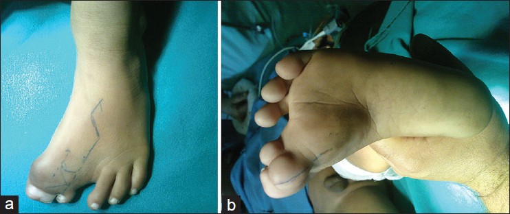
A 4-year-old baby brought to us with congenital anomaly of the left foot and difficulty in wearing footwear. There was also history of ulceration when closed shoes tried. No history any numbness, swelling over the sacrum, or any polysyndactyly of the other toes or fingers were present [Figure 1]. | Figure 1: (a and b) Preoperative marking with Z-plasty planned over the dorsum and fillet incision planned over the ventral aspect
Click here to view |
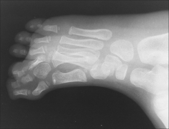
On examination, polysyndactyly of the great toe complete complex and nail duplication was seen. Pulses and sensations were normal and lateral/medial toe was dominated with restricted movements of the first phalangeal joint. Joint was normal except for slight out turning of the forefoot while walking. Ingrowing toe nail appearance was present. X-ray showed triple phalanx showing symphalangism and duplication of the metacarpophalangeal architecture [Figure 2]. | Figure 2: X-ray foot revealed the triple phalanx showing symphalangism and duplication of the metacarpophalangeal architecture duplication; symphalangyeasm of first ray; triplation of the phalanges of first toe
Click here to view |
Operative plan revealed disarticulation of the joint after filling of the extra bony architecture done and centralization of the tendon strips done over the dominant toe after placing an axial K-wire [Figure 3]. Flaps were refashioned and closure done over suction drains. Splintage was applied [Figure 4]. Dressing was opened in seventh postoperative period and skin was healthy. K-wire was removed after 6 weeks of surgery. | Figure 4: X-ray showing results achieved after amputation of the extra ray
Click here to view |
 Discussion Discussion | |  |
Polydactyly is a common congenital anomaly of the foot classified as preaxial, central, or postaxial depending on the location of the duplication. Approximately, 15% of all duplications of the toes are preaxial. [6] The incidence of polydactyly has been reported to be 1 per 1,000 live births. [4]
Polydactyly of the foot has been classified according to anatomic differences in bony structures, external appearance of involved digits, and associated anomalies. A new subtype of the metatarsal type of the medial ray (preaxial) polydactyly of the foot is presented. This includes triplication of the first metatarsal triplication of the tendons and three separate big toes. [7] There have been many works on the classification of polydactyly, but a great diversity of the phenotypes makes it difficult to categorize polydactyly clearly. Swanson suggested a classification with criteria based on the position of the extra digit on the hand or foot. [8] When polydactyly affects the thumb or the great toe, it is classified as radial/tibial/preaxial polydactyly; when it appears on the little finger, the polydactyly is referred to as ulnar/fibular/postaxial. If the three central digits are affected, the condition is referred to as central polydactyly. Because this classification has no confusion between the morphologic axis and the metapterygial axis, it is more reliable than other classification systems. The coexistence of preaxial and postaxial polydactyly of the hand and the foot crossed polydactyly. [4]
Polydactyly has been regarded as a hereditary malformation and is usually inherited as an autosomal dominant trait, as are most isolated malformations of the limbs. [4] The genes responsible for limb malformations are characterized by low penetrance and extreme variability. In fact, preaxial and postaxial polydactyly are inherited together. Advances in molecular biology techniques have enabled the identification of gene loci that are responsible for human polydactyly phenotypes. Some genes are also found to be related to the development of polydactyly, such as Greig cephalopolysyndactyly syndrome (GLI3) and sonic hedgehog (SHH). GLI3 plays a major role in early limb development and a point mutation in the GLI gene results in various types of polydactyly. GLI3-associated polydactyly has a phenotype of nonmirror images. Conversely, polydactyly caused by mutations in the SHH gene is triphalangeal and shows mirror image digits. In addition, fibroblast growth factor (FGF), homeobox D (5'-HoxD), and homeobox protein aristaless-like 4 (ALX4) are also known to be involved in the pathogenesis of polydactyly. In this case, although we did not perform a mutation analysis in the infant, biphalangeal polydactyly and nonmirror images of both feet suggest the possibility of a mutation of GLI3. [4]
Surgical correction of preaxial polydactyly is generally more complex with poor long-term results. [5] Complications include recurrent hallux varus, splaying of the first ray and a short first metatarsal that does not bear adequate weight. Treatment of central ray duplication is not well-publicized because of its rare presentation. In most cases, supernumerary central digits can be excised through a racquet-shaped incision. Treatment ranges from shoe modification to complex surgical procedures. [9] We have achieved the full success after surgery and child is having normal movements of the toes and foot.
 Conclusion Conclusion | |  |
General principles recommend saving the digit that is the most developed, that has the most normal metatarsophalangeal articulation and that will give the best contour to the foot. Surgery should not be delayed much beyond walking age to allow the maximum time for the bones to remodel. Nevertheless, surgery can be performed at any age as in our series with good results. Management of polydactyly of the foot may appear simple at first glance, but the multiformity of its configuration deserves careful consideration before and during surgical correction. Whatever the motive for the patient to consult, shoe problems, pain, or cosmetic reasons, the treatment should be individualized. If surgical correction is elected, it should lead to proper alignment of toes and comfort in wearing shoes. In case of polydactyly, familial transmission should be kept in mind and in future investigated for prenatal diagnosis.
 References References | |  |
| 1. | Biere SS, Lagarde SM, Wust AF, Steller EP. An unusual case of polydactyly. Orthopedics Orthopedics 2009;32: pii. 
|
| 2. | Muneuchi G, Kogure T, Sano N, Hamamoto Y, Kishikawa Y, Tamai M, et al. Rubinstein-Taybi syndrome (RTS) with postaxial polydactyly of the foot: 4-year follow-up until improvement of dysbasia. Congenit Anom 2005;45:65-6. 
|
| 3. | Turra S, Gigante C, Bisinella G. Polydactyly of the foot. J Pediatr Orthop B 2007;16:216-20. 
|
| 4. | Zun KH, Kim MW, Choi HM. Crossed polydactyly prenatally diagnosed by 2- and 3-dimensional sonography. J Ultrasound Med 2007;26:529-34. 
|
| 5. | Rayan GM, Frey B. Ulnar polydactyly. Plast Reconstr Surg 2001;107:1449-54. 
|
| 6. | Belthur MV, Linton JL, Barnes DA. The spectrum of preaxial polydactyly of the foot. J Pediatr Orthop 2011;31:435-47. 
|
| 7. | Akin S, Özcan M. A non-classified preaxial polydactyly of the foot. European J Plast Surg 1997;20:161-3. 
|
| 8. | Swanson AB. A classification for congenital limb malformations. J Hand Surg 1976;1:8-22. 
|
| 9. | Galois L, Mainard D, Delagoutte JP. Polydactyly of the foot. Literature review and case presentations. Acta Orthop Belg 2002;68:376-80. 
|
[Figure 1], [Figure 2], [Figure 3], [Figure 4]
|


