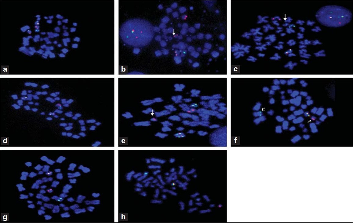
Figure 1: (a) D-FISH with LSI PML– RARA on metaphase cell shows normal PML allele (red signal), normal RARA allele (green signal) reciprocal PML– RARA fusion on der(15) (yellow signal) and der(17) (yellow signal). (b) LSI PML– RARA on metaphase cell shows PML– RARA fusion on der(15) (yellow signal) and residual PML on der(17) (red signal) (white arrow). (c) LSI PML– RARA on metaphase cell shows PML– RARA fusion on der(15) (yellow signal), also shows Aqua CEP 17 normal and der(17) (white arrow). (d) LSI PML– RARA on metaphase cell shows PML– RARA fusion on der(15) (yellow signal) and duplication of PML– RARA on i(17q) (yellow signals). (e) LSI PML– RARA on metaphase cell shows PML– RARA fusion on der (15) (yellow signal), residual RARA on der(15) (green signal) next to PML– RARA fusion and residual PML signal on der(17) (red signal) (white arrow). (f) Dual color RARA break-apart probe on metaphase cell shows normal RARA allele on 17 (yellow signal), residual RARA on der(17) (red signal) and residual RARA on der(11) at band 11q23 (green signal). (g) Dual color RARA break-apart probe on metaphase cell shows normal RARA allele on 17 (yellow signal), residual RARA on der17 (red signal) and residual RARA on der(11) at band 11q13 (green signal). (h) Dual color RARA break-apart probe on metaphase cell shows normal RARA allele on 17 (yellow signal), residual RARA on der(17) (red signal) and residual RARA on der(2) at band 2p21 (green signal)