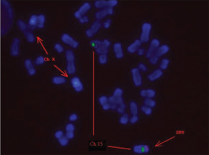
Figure 3: Fluorescence in situ hybridization on metaphase chromosomes using CEP X/SRY specific probes. Arrows indicate two X chromosome centromeric signals (blue) and one SRY specific signal (red) on the ends of chromosome 15. Fluorescent in situ hybridization (FISH) shows fluorescence illumination of the long arm of the Y chromosome and the short arm (q11 of the Y chromosome) of the derivative chromosome 15. The images were obtained separately and were captured. FISH result showed that there were two chromosomes X and one derived chromosome 15 contained Yq11.1 signals in peripheral blood lymphocyte from the patient.