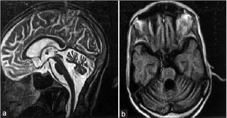
Figure 2: (a) MRI of 40 years old SCA1 patient II9. Strictly mid-sagittal section, showing normal Fundus and diffused cerebellar atrophy and preserved brainstem, (b) Image on axial section through the pons and the cerebellum. The cerebellar vermis and cortex are atrophied