
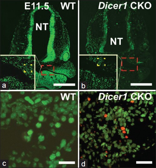
Figure 7: EdU (green) and ApopTag (red) staining in coronal sections of E11.5 Dicer1 CKO. a, c. WT. b, d. Dicer1 CKO. High magnification of dotted boxes shown in white inset box. NT: neural tube. Bar = 500 μm (a, b); 20μm (C, D)
|
|
Close |
|
Figure 7: EdU (green) and ApopTag (red) staining in coronal sections of E11.5 Dicer1 CKO. a, c. WT. b, d. Dicer1 CKO. High magnification of dotted boxes shown in white inset box. NT: neural tube. Bar = 500 μm (a, b); 20μm (C, D)
|
|