Indian J Med Microbiol Close
 

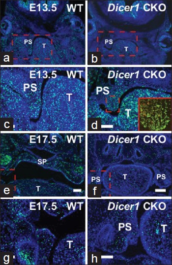
Figure 8: EdU (green) and ApopTag (red) staining at later time points. Right. WT. Left. Dicer1 CKO. Inset (d) highlights apoptosis. Boxes in (a, b), (e, f) shown in (c, d), (g, h), respectively. DAPI (blue). T: tongue; PS: palatal shelves; SP: secondary palate. Bar = 500μm

Figure 8: EdU (green) and ApopTag (red) staining at later time points. Right. WT. Left. <i>Dicer1</i> CKO. Inset (d) highlights apoptosis. Boxes in (a, b), (e, f) shown in (c, d), (g, h), respectively. DAPI (blue). T: tongue; PS: palatal shelves; SP: secondary palate. Bar = 500μm