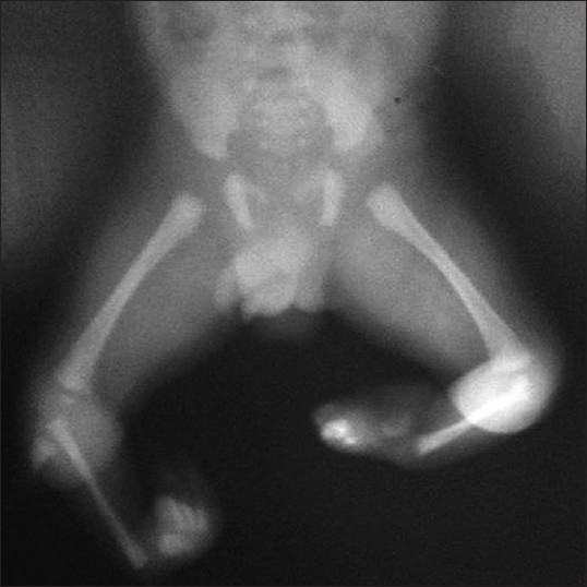
Figure 2: X - ray showing normal hip joint, normal lower end of femur, complete absence of both tibiae with small cartilaginous anlage, and presence of fibula on both legs. Both the right and left foot had three tarsal bones, two metatarsals, and three toes each having two phalanges each