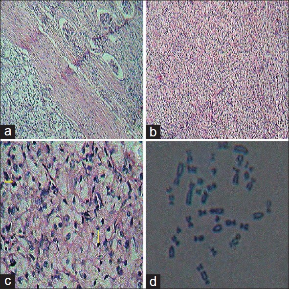
Figure 2: Immunohistochemistry of clear cell renal carcinoma. (a) Low power (×10) view of clear cell renal carcinoma with few glomeruli adjacent to the lesion. (b) Low power (×10) view of clear cell renal carcinoma. (c) High power (×40) of clear cell renal carcinoma comprised of round oval to polygonal cells with mildly pleomorphic vesicular nuclei, inconspicuous nucleoli and clear cytoplasm. Increased mitosis (shown in yellow arrow) was observed. (d) Karyotyping analysis of Von Hippel-Lindau gene mutation with clear cell renal carcinoma