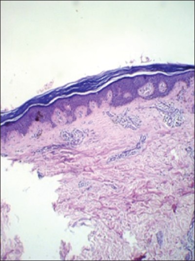
Figure 3: Histopathology of the skin lesion shows epidermis was mildly atrophic with hyperkeratosis. Basal layers showed mild vacuolar change, papillary dermis with melanin incontinence, and dilated dermal vessels (hematoxylin and eosin (H and E, ×10)