
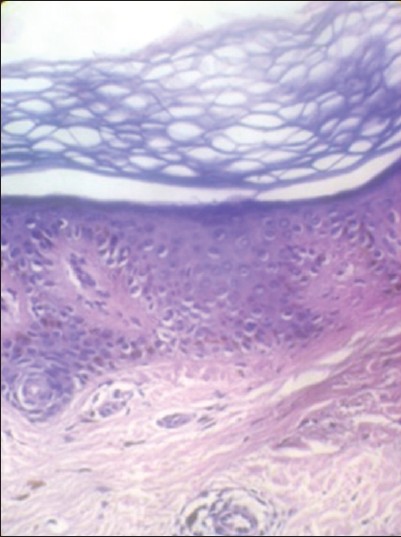
Figure 4: Histopathology of the skin lesion: In high power, section shows increase in pigment extending into the stratum spinosum. Dermis showed mild perivascular and periadnexeal lymphomononuclear infitrate (H and E, × 10)
|
|
Close |
|
Figure 4: Histopathology of the skin lesion: In high power, section shows increase in pigment extending into the stratum spinosum. Dermis showed mild perivascular and periadnexeal lymphomononuclear infitrate (H and E, × 10)
|
|