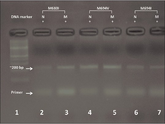
Figure 3: Deoxyribonucleic acid (DNA) electrophoresis of amplified segments of familial Mediterranean fever gene (~200 bp, upper arrow) using normal and mutant primers corresponding to each of the four studied mutations, the distal fragment is that of primer (bottom arrow). Lane 1 is a marker DNA, lanes 2, 3 heterozygous mutants M680I, lanes 4, 5 heterozygous mutant M694V and lanes 6, 7 heterozygous mutants M694I