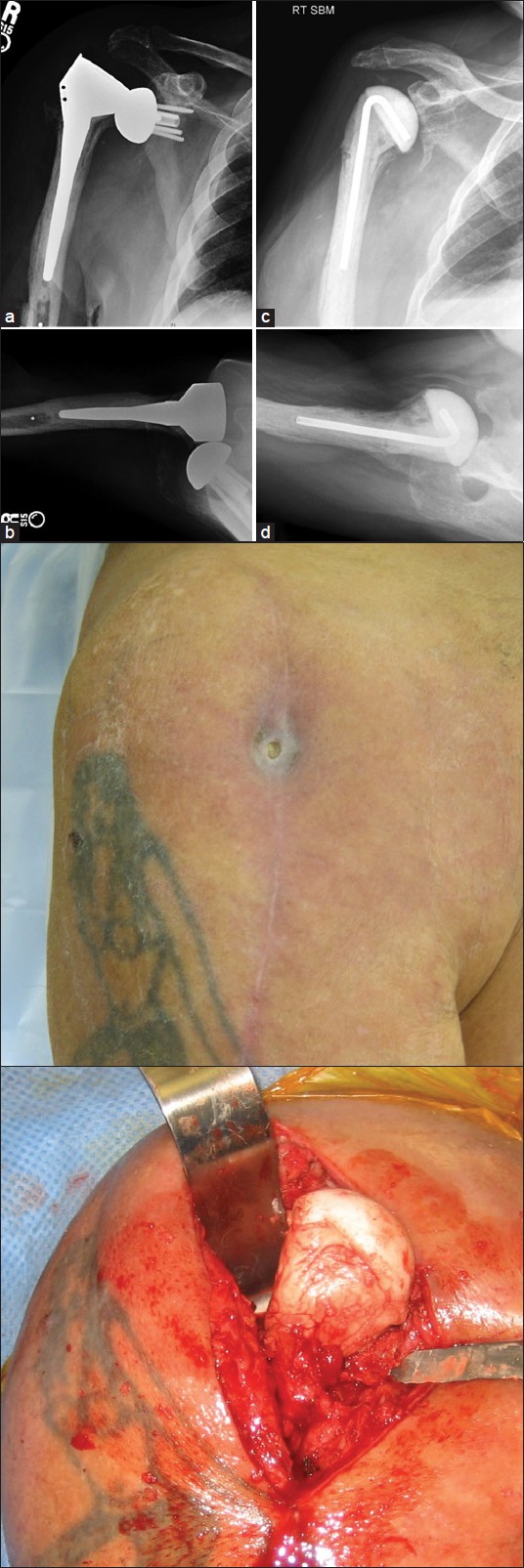
Figure 3: (a-d): An 80-year-old man (Case 5) presented with a draining sinus and a dislocated, infected reverse replacement 6 months after primary reverse replacement at an outside institution that was complicated with reoperation the same day after a recovery room dislocation. (a and b) Dislocation of reverse replacement. Glenosphere position is acceptable, but the entire humeral head was essentially resected, likely compromising stability. (c and d) Postoperative radiographs of preformed antibiotic spacer Figure 3e: Preoperative draining sinus Figure 3f: An intraoperative photograph after irrigation and debridement, removal of glenosphere, slot osteotomy and cementation of preformed antibiotic spacer (Exactech Interspace, Gainesville, FL, USA) with additional antibiotic-loaded cement. Notice how the humeral head is devoid of any soft tissue attachments creating a shoulder with profound anterosuperior escape