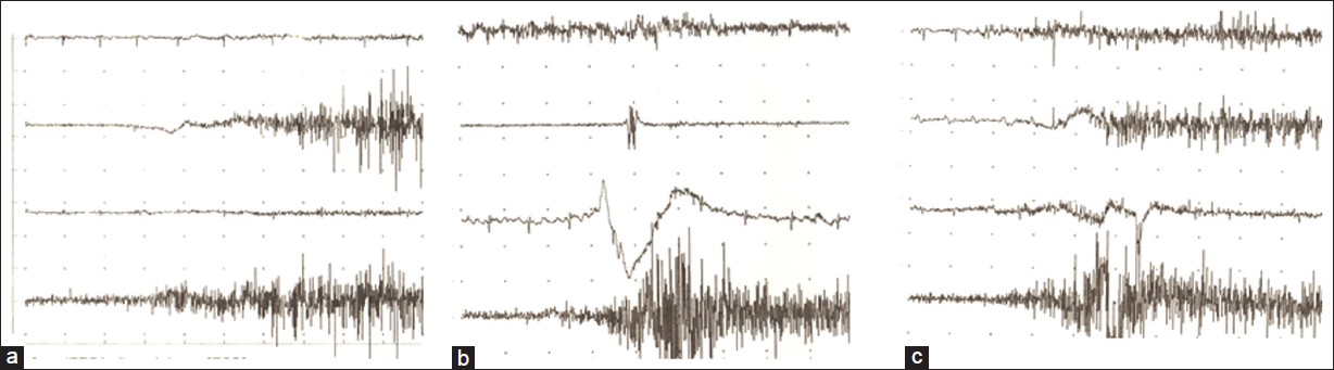
Figure 4: Patterns of abnormal electromyography activity. In trace, the four electromyographys shown in order from top to bottom are pectoralis major (PM), anterior deltoid (AD), latissimus dorsi (LD), and infraspinatus (IS). (a) Abnormal electromyography activity in AD during extension. (b) Abnormal electromyography activity in PM and LD during extension. (c) Abnormal electromyography activity in PM during external rotation